

What Is the Cause of Toxicity of Silicone Oil?
- PMID: 35009415
- PMCID: PMC8745808
- DOI: 10.3390/ma15010269
What Is the Cause of Toxicity of Silicone Oil?
Abstract
Purpose: To investigate the toxicity of the low-molecular-weight components (LMWCs) in ophthalmic silicone oils (SilOils) on retinal cell lines.
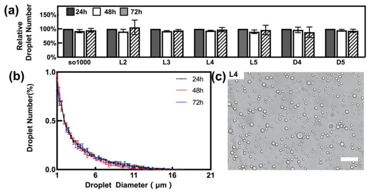
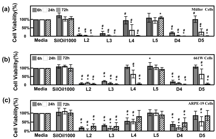
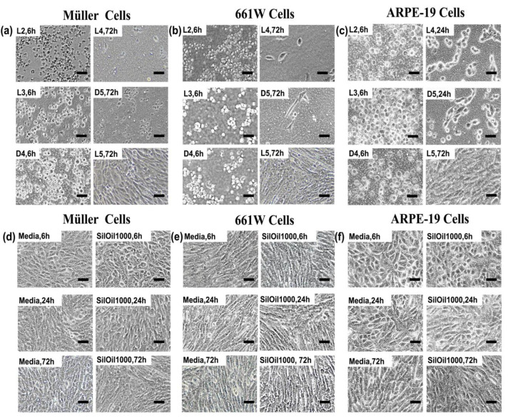
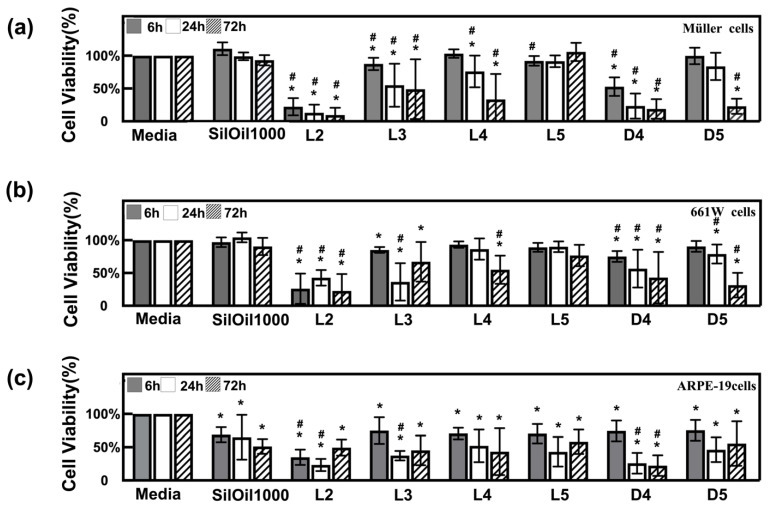
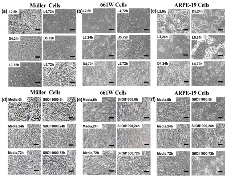
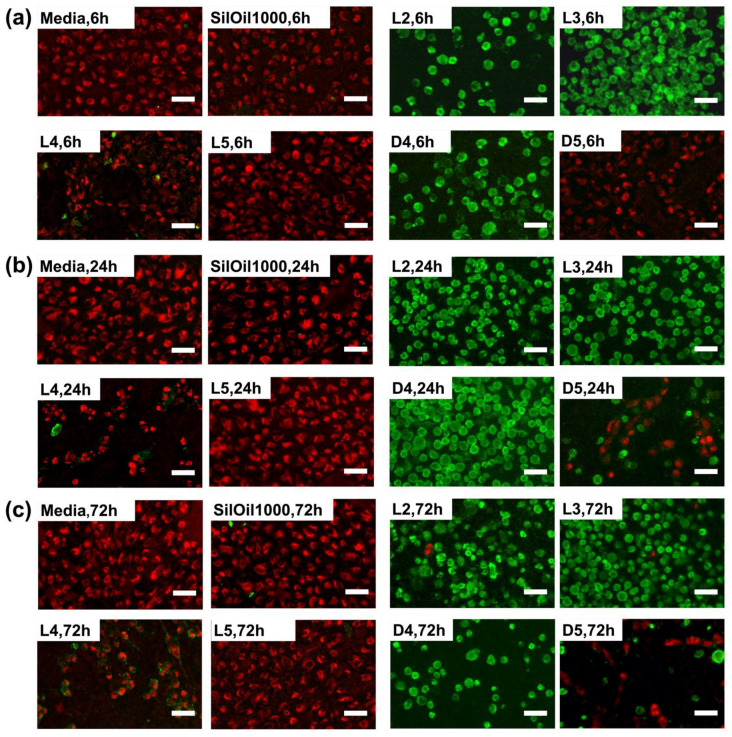
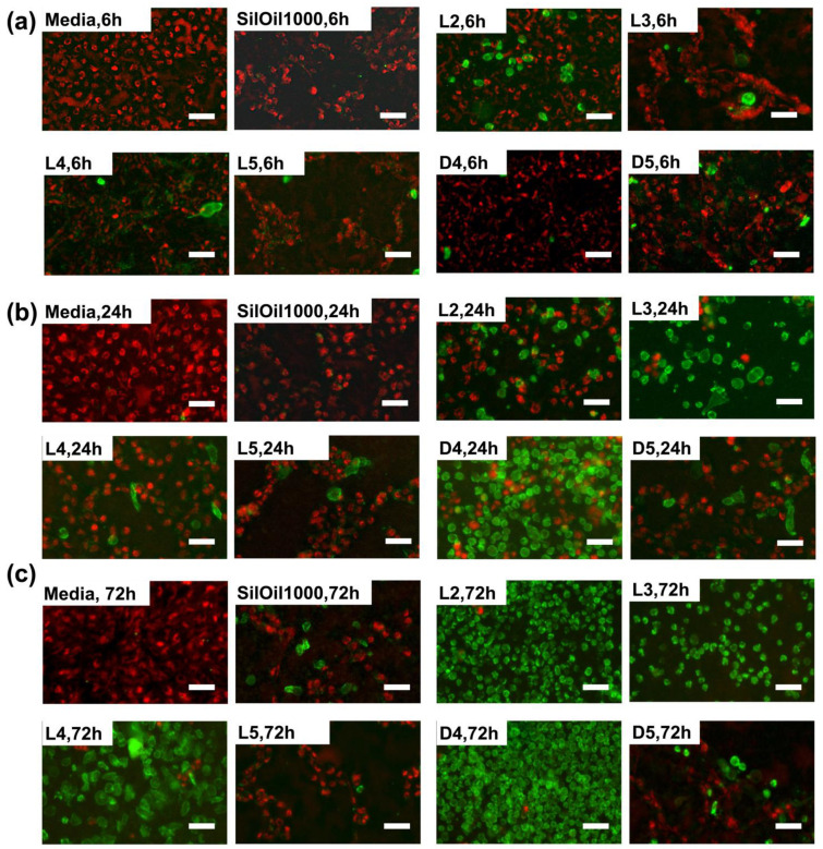
Methods: The toxicity of six types of LMWCs were studied and compared with conventional SilOil 1000 cSt. In vitro cytotoxic tests of LMWCs, in both liquid and emulsified forms, on three retinal cell lines (Müller cells (rMC-1), photoreceptor cells (661W) and retinal pigment epithelial cells (ARPE-19)) were conducted using a transwell cell culturing system. The morphology and viability of cells were assessed by light microscopy and Cell Counting Kit-8 (CCK-8) assay at different time points (6, 24 and 72 h). The ARPE-19 apoptotic pathway was investigated by Mitochondrial Membrane Potential/Annexin V Apoptosis Kit at different time points (6, 24 and 72 h).
Results: Apart from dodecamethylpentasiloxane (L5), all liquid LMWCs showed varying degrees of acute cytotoxicity on retinal cell lines within 72 h. Emulsified LMWCs showed comparable cytotoxicity with liquid LMWCs on retinal cell lines. Cyclic LMWCs, octamethylcyclotetrasiloxane (D4) and decamethylcyclopentasiloxane (D5) had significantly higher cytotoxicity when compared with their linear counterparts decamethyltetrasiloxane (L4) and L5 with similar molecular formula. Using ARPE-19 cells as an example, we showed that LMWCs induce the apoptosis of retinal cells.
Conclusions: Most LMWCs, in both liquid and emulsified forms, can induce acute cytotoxicity. In addition, cyclic LMWCs are suspected to have higher cytotoxicity than their linear counterparts. Therefore, LMWCs are suspected to be the main cause of the long-term toxicity of ophthalmic SilOil, due to their toxicity and propensity to cause ophthalmic SilOil to emulsify. The amount of LMWCs should be considered as the paramount parameter when referring to the quality of SilOil.
Keywords: Müller cell (rMC-1); emulsification; low-molecular-weight component (LMWC); photoreceptor cell (661W); polydimethylsiloxane; retinal toxicity.
Conflict of interest statement
All authors whose names are listed in this manuscript certify that they have no affiliations with or involvement in any organisation or entity with any financial interest (such as honoraria; educational grants; participation in speakers’ bureaus; membership, employment, consultancies, stock ownership, or other equity interest; and expert testimony or patent-licensing arrangements), or non-financial interest (such as personal or professional relationships, affiliations, knowledge or beliefs) in the subject matter or materials discussed in this manuscript.
Figures
Similar articles
-
Comparative Study of Chemical Composition, Molecular and Rheological Properties of Silicone Oil Medical Devices.Transl Vis Sci Technol. 2019 Sep 11;8(5):9. doi: 10.1167/tvst.8.5.9. eCollection 2019 Sep. Transl Vis Sci Technol. 2019. PMID: 31588374 Free PMC article.
-
Determination of siloxanes in silicone products and potential migration to milk, formula and liquid simulants.Food Addit Contam Part A Chem Anal Control Expo Risk Assess. 2012 Aug;29(8):1311-21. doi: 10.1080/19440049.2012.684891. Epub 2012 May 11. Food Addit Contam Part A Chem Anal Control Expo Risk Assess. 2012. PMID: 22575024
-
Safety of silicone oils as intraocular medical device: An in vitro cytotoxicity study.Exp Eye Res. 2020 May;194:108018. doi: 10.1016/j.exer.2020.108018. Epub 2020 Mar 21. Exp Eye Res. 2020. PMID: 32209320
-
Characterization and toxicology evaluation of low molecular weight chitosan on zebrafish.Carbohydr Polym. 2020 Jul 15;240:116164. doi: 10.1016/j.carbpol.2020.116164. Epub 2020 Apr 13. Carbohydr Polym. 2020. PMID: 32475540
-
Complications of emulsified silicone oil after retinal detachment repair.Semin Ophthalmol. 2014 Sep-Nov;29(5-6):312-8. doi: 10.3109/08820538.2014.962181. Semin Ophthalmol. 2014. PMID: 25325856 Review.
Cited by
-
Assessment of Silicone Oil Emulsification: A Comparison of Currently Applied Methods.J Ophthalmol. 2023 Apr 24;2023:8114530. doi: 10.1155/2023/8114530. eCollection 2023. J Ophthalmol. 2023. PMID: 37139082 Free PMC article.
-
Effect of silicone oil versus gas tamponade on macular layer microstructure after pars plana vitrectomy for macula on rhegmatogenous retinal detachment.BMC Ophthalmol. 2024 Mar 14;24(1):119. doi: 10.1186/s12886-024-03377-x. BMC Ophthalmol. 2024. PMID: 38486220 Free PMC article.
-
Retinal and Corneal Changes Associated with Intraocular Silicone Oil Tamponade.J Clin Med. 2022 Sep 5;11(17):5234. doi: 10.3390/jcm11175234. J Clin Med. 2022. PMID: 36079165 Free PMC article. Review.
-
Vitreous Substitutes from Bench to the Operating Room in a Translational Approach: Review and Future Endeavors in Vitreoretinal Surgery.Int J Mol Sci. 2023 Feb 7;24(4):3342. doi: 10.3390/ijms24043342. Int J Mol Sci. 2023. PMID: 36834754 Free PMC article. Review.
-
Emulsification of Silicone Oils: Altering Factors and Possible Complications-A Narrative Review.J Clin Med. 2024 Apr 20;13(8):2407. doi: 10.3390/jcm13082407. J Clin Med. 2024. PMID: 38673681 Free PMC article. Review.
References
Grants and funding
LinkOut - more resources
Full Text Sources
