

Validation of Visually Identified Muscle Potentials during Human Sleep Using High Frequency/Low Frequency Spectral Power Ratios
- PMID: 35009594
- PMCID: PMC8747095
- DOI: 10.3390/s22010055
Validation of Visually Identified Muscle Potentials during Human Sleep Using High Frequency/Low Frequency Spectral Power Ratios
Abstract
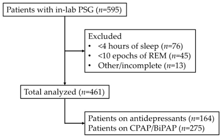
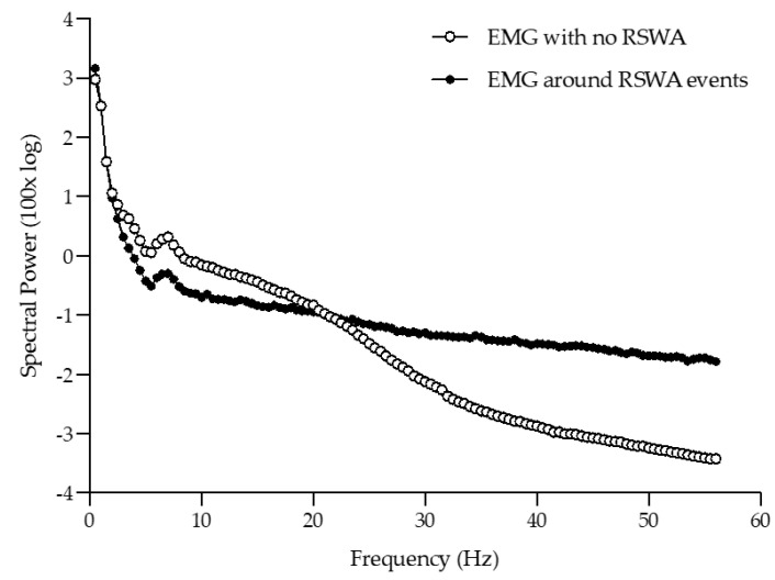
Surface electromyography (EMG), typically recorded from muscle groups such as the mentalis (chin/mentum) and anterior tibialis (lower leg/crus), is often performed in human subjects undergoing overnight polysomnography. Such signals have great importance, not only in aiding in the definitions of normal sleep stages, but also in defining certain disease states with abnormal EMG activity during rapid eye movement (REM) sleep, e.g., REM sleep behavior disorder and parkinsonism. Gold standard approaches to evaluation of such EMG signals in the clinical realm are typically qualitative, and therefore burdensome and subject to individual interpretation. We originally developed a digitized, signal processing method using the ratio of high frequency to low frequency spectral power and validated this method against expert human scorer interpretation of transient muscle activation of the EMG signal. Herein, we further refine and validate our initial approach, applying this to EMG activity across 1,618,842 s of polysomnography recorded REM sleep acquired from 461 human participants. These data demonstrate a significant association between visual interpretation and the spectrally processed signals, indicating a highly accurate approach to detecting and quantifying abnormally high levels of EMG activity during REM sleep. Accordingly, our automated approach to EMG quantification during human sleep recording is practical, feasible, and may provide a much-needed clinical tool for the screening of REM sleep behavior disorder and parkinsonism.
Keywords: EMG; Parkinson’s disease; RBD; REM sleep behavior disorder; REM sleep without atonia; electromyography; parkinsonism; polysomnography; spectral power.
Conflict of interest statement
The authors declare no conflict of interest.
Figures
Similar articles
-
Comparison between an automatic and a visual scoring method of the chin muscle tone during rapid eye movement sleep.Sleep Med. 2014 Jun;15(6):661-5. doi: 10.1016/j.sleep.2013.12.022. Epub 2014 Mar 5. Sleep Med. 2014. PMID: 24831249 Clinical Trial.
-
Night-to-night variability of automatic quantitative parameters of the chin EMG amplitude (Atonia Index) in REM sleep behavior disorder.J Clin Sleep Med. 2013 Mar 15;9(3):253-8. doi: 10.5664/jcsm.2490. J Clin Sleep Med. 2013. PMID: 23493642 Free PMC article.
-
Quantification of electromyographic activity during REM sleep in multiple muscles in REM sleep behavior disorder.Sleep. 2008 May;31(5):724-31. doi: 10.1093/sleep/31.5.724. Sleep. 2008. PMID: 18517042 Free PMC article.
-
Defining muscle activities for assessment of rapid eye movement sleep behavior disorder: from a qualitative to a quantitative diagnostic level.Sleep Med. 2013 Aug;14(8):729-33. doi: 10.1016/j.sleep.2012.09.028. Epub 2012 Dec 13. Sleep Med. 2013. PMID: 23245755 Review.
-
Precision Medicine in Rapid Eye Movement Sleep Behavior Disorder.Sleep Med Clin. 2019 Sep;14(3):351-362. doi: 10.1016/j.jsmc.2019.04.003. Epub 2019 Jun 21. Sleep Med Clin. 2019. PMID: 31375203 Review.
Cited by
-
Transforming Sleep Monitoring: Review of Wearable and Remote Devices Advancing Home Polysomnography and Their Role in Predicting Neurological Disorders.Biosensors (Basel). 2025 Feb 17;15(2):117. doi: 10.3390/bios15020117. Biosensors (Basel). 2025. PMID: 39997019 Free PMC article. Review.
References
-
- Berry R.B., Brooks R., Gamaldo C.E., Harding S.M., Marcus C.L., Vaughn B.V., Tangredi M.M. The AASM Manual for the Scoring of Sleep and Associated Events: Rules, Terminology and Technical Specifications. American Academy of Sleep Medicine; Darien, IL, USA: 2012. Version 2.0.
-
- Jouvet M., Michel M., Courjon J. Sur la mise en jeu de deux mechanismes a l’expression electro-encephalographique differente au cours du sommeil physiologique chez le Chat. Comptes Rendus Seances L’Academie Sci. 1959;248:3043–3045. - PubMed
MeSH terms
Grants and funding
LinkOut - more resources
Full Text Sources
