. 2021 Dec 23;12(1):33.
doi: 10.3390/nano12010033.

Lateral Flow Immunoassay with Quantum-Dot-Embedded Silica Nanoparticles for Prostate-Specific Antigen Detection

Affiliations

Lateral Flow Immunoassay with Quantum-Dot-Embedded Silica Nanoparticles for Prostate-Specific Antigen Detection

Sungje Bock et al. Nanomaterials (Basel). .

Abstract

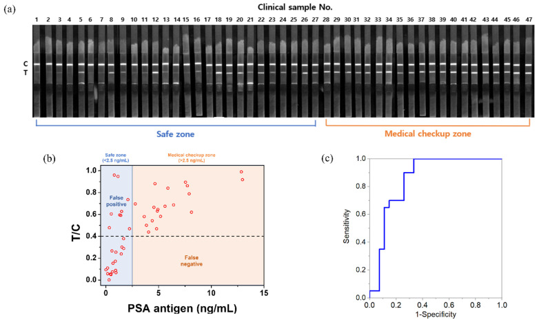
Prostate cancer can be detected early by testing the presence of prostate-specific antigen (PSA) in the blood. Lateral flow immunoassay (LFIA) has been used because it is cost effective and easy to use and also has a rapid sample-to-answer process. Quantum dots (QDs) with very bright fluorescence have been previously used to improve the detection sensitivity of LFIAs. In the current study, a highly sensitive LFIA kit was devised using QD-embedded silica nanoparticles. In the present study, only a smartphone and a computer software program, ImageJ, were used, because the developed system had high sensitivity by using very bright nanoprobes. The limit of PSA detection of the developed LFIA system was 0.138 ng/mL. The area under the curve of this system was calculated as 0.852. The system did not show any false-negative result when 47 human serum samples were analyzed; it only detected PSA and did not detect alpha-fetoprotein and newborn calf serum in the samples. Additionally, fluorescence was maintained on the strip for 10 d after the test. With its high sensitivity and convenience, the devised LFIA kit can be used for the diagnosis of prostate cancer.

Keywords: lateral flow immunoassay; prostate cancer; prostate-specific antigen; quantum dot; quantum-dot-embedded silica nanoparticles.


Conflict of interest statement

The authors declare no conflict of interest.

Figures

Figure 1
Figure 1
(a) Schematic illustration of QD2-PSA Ab fabrication (this figure is not drawn to scale); (b) transmission electron microscopy (TEM) images of QD2. The inset depicts individual QD2; (c) UV–Vis absorbance of SiO2 NPs, QD2, and QD2-PSA Ab; (d) PL intensity of SiO2 NPs, QD2, and QD2-PSA Ab; (e) PL intensity comparison between QDs and QD2.
Figure 2
Figure 2
(a) Schematic illustration of lateral flow immunoassay (LFIA) process and precise analysis; (b) fluorescence image showing only the red channel of the test strip with PSA developed under a 365 nm UV lamp; (c) T/C value and fitting curve of the fluorescence intensity generated for each concentration of PSA.
Figure 3
Figure 3
(a) Fluorescence image showing only the red channel of the test strip after the development of human serum samples under a 365 nm UV lamp; (b) T/C value and concentration of PSA of each human serum sample; (c) ROC curve.
Figure 4
Figure 4
(a) Fluorescence image showing only the red channel of the test strip after the development of PSA, alpha-fetoprotein (AFP), and newborn calf serum (NCS) under a 365 nm UV lamp for the selectivity test; (b) T/C value of PSA, AFP, and NCS in strip tests.
Figure 5
Figure 5
(a) Fluorescence image showing only the red channel of the test strip (stored for 10 d) after the development of 4.557 ng/mL of human serum sample under a 365 nm UV lamp for the stability test; (b) normalized T/C value of the developed human serum sample.

Similar articles

Cited by

References

    1. Siegel R.L., Miller K.D., Fuchs H.E., Jemal A. Cancer Statistics, 2021. CA Cancer J. Clin. 2021;71:7–33. doi: 10.3322/caac.21654. - DOI - PubMed
    1. Rawla P. Epidemiology of prostate cancer. World J. Oncol. 2019;10:63. doi: 10.14740/wjon1191. - DOI - PMC - PubMed
    1. Kasivisvanathan V., Rannikko A.S., Borghi M., Panebianco V., Mynderse L.A., Vaarala M.H., Briganti A., Budäus L., Hellawell G., Hindley R.G. MRI-targeted or standard biopsy for prostate-cancer diagnosis. N. Engl. J Med. 2018;378:1767–1777. doi: 10.1056/NEJMoa1801993. - DOI - PMC - PubMed
    1. Etzioni R., Tsodikov A., Mariotto A., Szabo A., Falcon S., Wegelin J., Karnofski K., Gulati R., Penson D.F., Feuer E. Quantifying the role of PSA screening in the US prostate cancer mortality decline. Cancer Causes Control. 2008;19:175–181. doi: 10.1007/s10552-007-9083-8. - DOI - PMC - PubMed
    1. You J., Cozzi P., Walsh B., Willcox M., Kearsley J., Russell P., Li Y. Innovative biomarkers for prostate cancer early diagnosis and progression. Crit. Rev. Oncol. Hematol. 2010;73:10–22. doi: 10.1016/j.critrevonc.2009.02.007. - DOI - PubMed