. 2021 Dec 23;14(1):42.
doi: 10.3390/nu14010042.

Lactoferrin Prevents Hepatic Injury and Fibrosis via the Inhibition of NF-κB Signaling in a Rat Non-Alcoholic Steatohepatitis Model

Affiliations

Lactoferrin Prevents Hepatic Injury and Fibrosis via the Inhibition of NF-κB Signaling in a Rat Non-Alcoholic Steatohepatitis Model

Yoshinaga Aoyama et al. Nutrients. .

Abstract

Non-alcoholic steatohepatitis (NASH) can cause liver cirrhosis and hepatocellular carcinoma (HCC), with cases increasing worldwide. To reduce the incidence of liver cirrhosis and HCC, NASH is targeted for the development of treatments, along with viral hepatitis and alcoholic hepatitis. Lactoferrin (LF) has antioxidant, anti-cancer, and anti-inflammatory activities. However, whether LF affects NASH and fibrosis remains unelucidated. We aimed to clarify the chemopreventive effect of LF on NASH progression. We used a NASH model with metabolic syndrome established using connexin 32 (Cx32) dominant negative transgenic (Cx32ΔTg) rats. Cx32ΔTg rats (7 weeks old) were fed a high-fat diet and intraperitoneally injected with dimethylnitrosamine (DMN). Rats were divided into three groups for LF treatment at 0, 100, or 500 mg/kg/day for 17 weeks. Lactoferrin significantly protected steatosis and lobular inflammation in Cx32ΔTg rat livers and attenuated bridging fibrosis or liver cirrhosis induced by DMN. By quantitative RT-PCR, LF significantly down-regulated inflammatory (Tnf-α, Il-6, Il-18, and Il-1β) and fibrosis-related (Tgf-β1, Timp2, and Col1a1) cytokine mRNAs. Phosphorylated nuclear factor (NF)-κB protein decreased in response to LF, while phosphorylated JNK protein was unaffected. These results indicate that LF might act as a chemopreventive agent to prevent hepatic injury, inflammation, and fibrosis in NASH via NF-κB inactivation.

Keywords: NASH; connexin; fibrosis; hepatocarcinogenesis; lactoferrin.


Conflict of interest statement

The authors declare that they have no conflict of interest.

Figures

Figure 1
Figure 1
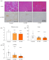
Preventive effect of lactoferrin on nonalcoholic steatohepatitis in rats. Connexin 32 dominant negative transgenic (Cx32ΔTg) rats were fed a high-fat diet (HFD), given an intraperitoneal injection of dimethylnitrosamine (DMN), and treated with lactoferrin (LF) for 17 weeks. (a) Representative histological findings of hematoxylin and eosin (H&E) stains in liver sections taken from Control, LF 100 mg/kg/day (LF100) or LF 500 mg/kg/day (LF500) rat groups. (be) Histopathological analysis of non-alcoholic steatohepatitis (NASH) was evaluated by severity scores for (b) steatosis, (c) lobular inflammation, (d) hepatocellular ballooning, and (e) a non-alcoholic fatty liver disease activity score (NAS). Data is shown as the mean ± SD, n = 15–16 per group, ** p < 0.01, *** p < 0.001, **** p < 0.0001 compared to the Control group.
Figure 2
Figure 2
Attenuation effect of lactoferrin on fibrosis in rat nonalcoholic steatohepatitis. Connexin 32 dominant negative transgenic (Cx32ΔTg) rats were fed a high-fat diet (HFD), given an intraperitoneal injection of dimethylnitrosamine (DMN), and treated with lactoferrin (LF) for 17 weeks. (a) Azan staining (upper panels) and α-smooth muscle actin (α-SMA; lower panels) immunohistochemical stains of liver sections from Control, LF 100 mg/kg/day (LF100), or LF 500 mg/kg/day (LF500) rat groups. (b) Azan staining was used to evaluate the fibrosis score and (c) percentage of fibrosis area. (d) α-SMA–positive area. Data is shown as the mean ± SD, n = 15–16 per group, *** p < 0.001, **** p < 0.0001 compared to the Control group.
Figure 3
Figure 3
Effect of lactoferrin on hepatocarcinogenesis in rat nonalcoholic steatohepatitis. Connexin 32 dominant negative transgenic (Cx32ΔTg) rats were fed a high-fat diet (HFD), given an intraperitoneal injection of dimethylnitrosamine (DMN), and treated with lactoferrin (LF) for 17 weeks. (a) Liver sections showing representative foci positive for glutathione S-transferase placental form (GST-P) from Control, LF 100 mg/kg/day (LF100), or LF 500 mg/kg/day (LF500) rat groups. (b) The number and (c) area of GST-P–positive hepatic foci. Data is shown as the mean ± SD, n = 15–16 per group.
Figure 3
Figure 3
Effect of lactoferrin on hepatocarcinogenesis in rat nonalcoholic steatohepatitis. Connexin 32 dominant negative transgenic (Cx32ΔTg) rats were fed a high-fat diet (HFD), given an intraperitoneal injection of dimethylnitrosamine (DMN), and treated with lactoferrin (LF) for 17 weeks. (a) Liver sections showing representative foci positive for glutathione S-transferase placental form (GST-P) from Control, LF 100 mg/kg/day (LF100), or LF 500 mg/kg/day (LF500) rat groups. (b) The number and (c) area of GST-P–positive hepatic foci. Data is shown as the mean ± SD, n = 15–16 per group.
Figure 4
Figure 4
Down-regulation of inflammatory cytokines by lactoferrin in rat nonalcoholic steatohepatitis. Connexin 32 dominant negative transgenic (Cx32ΔTg) rats were fed a high-fat diet (HFD), given an intraperitoneal injection of dimethylnitrosamine (DMN), and treated with lactoferrin (LF) for 17 weeks. (a) NormFinder stability values for candidate housekeeping genes (Gapdh, B2m, Actb, Ppia, and Gusb). (b,c) mRNA levels of (b) pro-inflammatory cytokines (Tnf-α, Il-6, Il-18, Ifn-γ, and Il-1β) and (c) pro-fibrotic cytokines (Tgf-β1, Timp1, Timp2, Col1a1, and Ctgf) in Control, LF 100 mg/kg/day (LF100), or LF 500 mg/kg/day (LF500) rat groups were measured using quantitative reverse transcription (RT)–PCR. Data is shown as the mean ± SD, n = 15–16 per group, * p < 0.05, ** p < 0.01, *** p < 0.001, **** p < 0.0001 compared to the Control group.
Figure 5
Figure 5
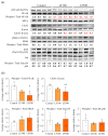
Down-regulation of inflammatory cytokines and deactivation of NF-κB and JNK signaling after the administration of lactoferrin in nonalcoholic steatohepatitis induced in Cx32 dominant negative transgenic rats. Connexin 32 dominant negative transgenic (Cx32ΔTg) rats were fed a high-fat diet (HFD), given an intraperitoneal injection of dimethylnitrosamine (DMN), and treated with lactoferrin (LF) for 17 weeks. (a) Protein levels of nuclear factor (NF)-κB-related (NF-κB, phosphorylated (p)NF-κB, IκB-α) and SAPK/JNK (Cdc42, Mkk4, pMkk4, Jnk, and pJnk) signaling proteins in Control, LF 100 mg/kg/day (LF100), or LF 500 mg/kg/day (LF500) rat groups were assessed by western blotting. Each lane represents a protein sample from an individual rat. Phospho, phosphorylated. (b) Data is shown as the mean ± SD. * p < 0.05, ** p < 0.01 compared to the Control group.

References

    1. Chalasani N., Younossi Z., Lavine J.E., Diehl A.M., Brunt E.M., Cusi K., Charlton M., Sanyal A.J. The diagnosis and management of non-alcoholic fatty liver disease: Practice guideline by the American Gastroenterological Association, American Association for the Study of Liver Diseases, and American College of Gastroenterology. Gastroenterology. 2012;142:1592–1609. doi: 10.1053/j.gastro.2012.04.001. - DOI - PubMed
    1. Younossi Z.M. Non-alcoholic fatty liver disease—A global public health perspective. J. Hepatol. 2019;70:531–544. doi: 10.1016/j.jhep.2018.10.033. - DOI - PubMed
    1. Younossi Z., Anstee Q.M., Marietti M., Hardy T., Henry L., Eslam M., George J., Bugianesi E. Global burden of NAFLD and NASH: Trends, predictions, risk factors and prevention. Nat. Rev. Gastroenterol. Hepatol. 2018;15:11–20. doi: 10.1038/nrgastro.2017.109. - DOI - PubMed
    1. Anstee Q.M., Targher G., Day C.P. Progression of NAFLD to diabetes mellitus, cardiovascular disease or cirrhosis. Nat. Rev. Gastroenterol. Hepatol. 2013;10:330–344. doi: 10.1038/nrgastro.2013.41. - DOI - PubMed
    1. Evans W.H., Martin P.E. Gap junctions: Structure and function (Review) Mol. Membr. Biol. 2002;19:121–136. doi: 10.1080/09687680210139839. - DOI - PubMed

MeSH terms