. 2021 Dec 22;12(1):18.
doi: 10.3390/ani12010018.

Evaluation of High-Volume Injections Using a Modified Dorsal Quadratus Lumborum Block Approach in Canine Cadavers

Affiliations

Evaluation of High-Volume Injections Using a Modified Dorsal Quadratus Lumborum Block Approach in Canine Cadavers

André Marchina-Gonçalves et al. Animals (Basel). .

Abstract

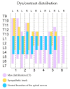
The quadratus lumborum (QL) block targets the fascial plane surrounding the QL muscle providing abdominal somatic and visceral analgesia. The extension of its analgesic effects is a subject of research, as it could not cover areas of the cranial abdomen in dogs. This study assesses in eight thawed canine cadavers, the distribution of high-volume injections (0.6 mL kg-1 of a mixture of methylene blue and iopromide) injected between the psoas minor muscle and the vertebral body of L1. Anatomical features of the area of interest were studied in two cadavers. In another six dogs, QL blocks were performed bilaterally under ultrasound-guidance. The distribution of contrast was evaluated by computed tomography (CT). Hypaxial abdominal muscles were dissected to visualize the dye spread (spinal nerves and sympathetic trunk) in 5 cadavers. The remaining cadaver was refrozen and cross-sectioned. CT studies showed a maximum distribution of contrast from T10 to L7. The methylene blue stained T13 (10%), L1 (100%), L2 (100%), L3 (100%), L4 (60%) and the sympathetic trunk T10 (10%), T11 (20%), T12 (30%), T13 (70%), L1 (80%), L2 (80%), L3 (60%) and L4 (30%). These findings may suggest that despite the high volume of injectate administered, this modified QL block could not produce somatic analgesia of the cranial abdomen, although it could provide visceral analgesia in dogs.

Keywords: abdominal analgesia; canine; locoregional anesthesia; ultrasound.


Conflict of interest statement

The authors declare no conflict of interest.

Figures
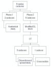
Figure 1
Figure 1
Schematic diagram of the experimental design. CT, computed tomography; QL, quadratus lumborum.
Figure 2
Figure 2
Ultrasound-guided approach to perform the QL block in dogs. Animals were positioned in lateral recumbency, and the transducer placed parallel to the last rib. The needle was inserted “in-plane” at the L1 level. QL, quadratus lumborum, L1; first lumbar vertebra, Cr, cranial; Cd, caudal; V, ventral; D, dorsal.
Figure 3
Figure 3
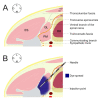
Schematic illustration of injection area at the L1 region in a dog placed in left lateral recumbency. (A) Main anatomical structures. (B) Final position of the needle and spreading of the injectate as observed in most hemiabdomens. L1, first lumbar vertebra; EO, external oblique muscle; IO, internal oblique muscle; TA, transverse abdominal muscle; ES, erector spinae muscles; QL, quadratus lumborum muscle; PM, psoas minor muscle; RK, right kidney; L, lateral; M, medial; D, dorsal; V, ventral.
Figure 4
Figure 4
Ultrasound images of the modified approach to the QL block. (A) Sonoanatomy of the L1 region. (B) Schematic superimposition of the main anatomical structures. (C) Final position of the needle tip between the vertebral body and the psoas minor muscle. (D) Distribution of the injectate. L1, first lumbar vertebra; EO, external oblique muscle; IO, internal oblique muscle; TP, transverse process; TAp, transverse abdominal aponeurosis; TA, transverse abdominal muscle; ES, erector spinae muscles; QL, quadratus lumborum muscle; IP, interfascial plane; PM, psoas minor muscle; VB, vertebral body; DI, distribution of injectate; L, lateral; M, medial; D, dorsal; V, ventral.
Figure 5
Figure 5
Computed tomographic images of the contrast spread after the administration of 0.6 mL kg−1 of a mixture of methylene blue and iopromide by a modified QL approach. (A) Volume-rendered three-dimensional projection image viewed from a ventral perspective of the thoracolumbar region. (B) Transverse image at L2 level with bone window setting. (C) Transverse image at L3 level showing contrasts presence in the left transverse abdominal plane. QL, quadratus lumborum block; L2; second lumbar vertebra; L3, third lumbar vertebra; Cr, cranial; Cd, caudal; L, left; R, right; D, dorsal; V, ventral.
Figure 6
Figure 6
Staining of the ventral branches of the spinal nerves evaluated by computed tomographic and anatomical dissection after the administration of 0.6 mL kg−1 of a mixture of methylene blue and iopromide by a modified QL approach. QL, quadratus lumborum block; L, left hemiabdomen; R, right hemiadbomen.
Figure 7
Figure 7
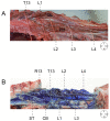
(A) Anatomical dissection of the thoracolumbar region. (B) Thoracolumbar region staining after methylene blue administration. T13; L1; L2; L3; L4, ventral branches of T13, L1, L2, L3 and L4 nerves respectively; QL, quadratus lumborum muscle; PM, psoas minor muscle; R13, 13th rib; ST, sympathetic trunk; CB, communicating branch; L, lateral; M, medial; Cr, cranial; Cd, caudal.
Figure 8
Figure 8
(A) Distribution of the dye through the quadratus lumborum and psoas minor muscles and spinal nerves. (B) Distribution of the dye through the lumbar sympathetic trunk after removing the right psoas minor muscle and the caudal aorta. T13; L1; L2; L3; L4, ventral branches of T13, L1, L2, L3 and L4 nerves respectively; GF, genitofemoral nerve; ST, sympathetic trunk; Ao, aorta; QL, quadratus lumborum muscle; PM, psoas minor muscle; Cr, cranial; Cd, caudal; R, right; L, left.
Figure 9
Figure 9
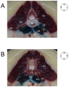
(A) Cranial view of a cross-section of the intervertebral disk and the caudal segment of L2. On the right side a spinal nerve can be observed leaving the intervertebral foramen and passing through the quadratus lumborum muscle. On the opposite side the presence of dye between the quadratus lumborum, the psoas minor muscles and the vertebral body can be observed. An intense coloration of the sympathetic trunk area between both psoas minor muscles is observed. (B) Dye distribution through the muscles and the sympathetic trunk area in a cranial vision of the center of L2. At this point, the spinal nerves cannot be seen. ES, erector spinae muscles; TP, transverse process; QL, quadratus lumborum muscle; PM, psoas minor muscle; ID, invetervebral disk; Ao, aorta; VC, vena cava; SN, spinal nerve; ND, nerve dyed; VB, vertebral body; ST, sympathetic trunk; D, dorsal; V, ventral; L, left side; R, right side.

References

    1. Ueshima H., Otake H., Lin J.A. Ultrasound-guided quadratus lumborum block: An updated review of anatomy and techniques. BioMed Res. Int. 2017;2017:2752876. doi: 10.1155/2017/2752876. - DOI - PMC - PubMed
    1. Garbin M., Portela D.A., Bertolizio G., Garcia-Pereira F., Gallastegui A., Otero P.E. Description of ultrasound-guided quadratus lumborum block technique and evaluation of injectate spread in canine cadavers. Vet. Anaesth. Analg. 2020;47:249–258. doi: 10.1016/j.vaa.2019.12.005. - DOI - PubMed
    1. Garbin M., Portela D.A., Bertolizio G., Gallastegui A., Otero P.E. A novel ultrasound-guided lateral quadratus lumborum block in dogs: A comparative cadaveric study of two approaches. Vet. Anaesth. Analg. 2020;47:810–818. doi: 10.1016/j.vaa.2020.08.003. - DOI - PubMed
    1. Alaman M., Bonastre C., de Blas I., Gomez-Alvarez C.M., Laborda A. Description of a novel ultrasound-guided approach for a dorsal quadratus lumborum block: A canine cadaver study. Vet. Anaesth. Analg. 2021 doi: 10.1016/j.vaa.2021.09.002. - DOI - PubMed
    1. Viscasillas J., Terrado J., Marti-Scharfhausen R., Castiñeiras D., Esteve V., Clancy N., Redondo J.I. A Modified Approach for the Ultrasound-Guided Quadratus Lumborum Block in Dogs: A Cadaveric Study. Animals. 2021;11:2945. doi: 10.3390/ani11102945. - DOI - PMC - PubMed