Review
. 2021 Dec 30;27(1):210.
doi: 10.3390/molecules27010210.

The Structural Basis of Peptide Binding at Class A G Protein-Coupled Receptors

Affiliations
Review

The Structural Basis of Peptide Binding at Class A G Protein-Coupled Receptors

Oanh Vu et al. Molecules. .

Abstract

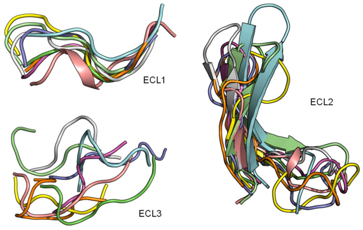
G protein-coupled receptors (GPCRs) represent the largest membrane protein family and a significant target class for therapeutics. Receptors from GPCRs' largest class, class A, influence virtually every aspect of human physiology. About 45% of the members of this family endogenously bind flexible peptides or peptides segments within larger protein ligands. While many of these peptides have been structurally characterized in their solution state, the few studies of peptides in their receptor-bound state suggest that these peptides interact with a shared set of residues and undergo significant conformational changes. For the purpose of understanding binding dynamics and the development of peptidomimetic drug compounds, further studies should investigate the peptide ligands that are complexed to their cognate receptor.

Keywords: class A GPCR; non-canonical amino acids; peptide GPCR; peptide docking.


Conflict of interest statement

The authors declare no conflict of interest.

Figures

Figure 1
Figure 1
Overview of nine co-crystal structures of class A peptide-GPCR. (Left) Comparison of peptide binding modes and crystallized peptides DAMGO (cyan), PMX53 (magenta), sAngII (beige), apelin derivative (salmon), ET-1 (green), gp120 (orange), vMIP-II (yellow), CX3CL1 (red), and 5P7-CCL5 (blue) at the receptors µ opioid receptor (µOR) (PDB ID: 6DDE), complement component 5a receptor (C5aR) (PDB ID: 6C1R), angiotensin II type 2 receptor (AT2R) (PDB ID: 5XJM), apelin receptor (APJR) (PDB ID: 5VBL), endothelin B receptor (ET-B) (PDB ID: 5GLH), C-C chemokine receptor type 5 (CCR5) (PDB ID: 6MEO), US28 (PDB ID: 4XT1), CXC-chemokine receptor 4 (CXCR4) (PDB ID: 4RWS), and CCR5 (PDB ID: 5UIW), respectively [22,32,34,37,38,39,40,41,42]. All receptors were aligned in the transmembrane region. The approximated extracellular border of the transmembrane region is marked in the upper dotted dark blue lines. The membrane region of GPCR receptors was calculated using the PPM server [65]. The lower blue bars and texts illustrate the depth of penetration for each peptide ligand. (Right) Classification tree of eight class A GPCRs with their nine peptide ligands in those nine listed structures.
Figure 2
Figure 2
Despite the diversity in the peptide engagement, their overlapping region at the core of their binding pocket suggests common ligand-GPCR interactions. (A) Superimposition of the nine peptides/class A GPCR complexes. (B) Overlay of all peptide ligands and zoom-in of the peptide region at the cores of GPCRs.
Figure 3
Figure 3
Rearrangements in the extracellular domain of peptide-activated GPCRs for peptide binding. (A) In the apo ET-B receptor (grey, PDB ID 5GLI), the N-terminus (orange) is lying over the ligand binding pocket. In the ET-1-bound state (cyan, PDB ID 5GLH), the bound ET-1 ligand (magenta) occupies the space of the N-terminus leading to its displacement [55]. (B) The crystal structure of antagonist-bound Y1 receptor (grey, PDB ID 5ZBQ) is also found with the N-terminus (orange) lying over the ligand binding pocket. The modeled peptide-bound Y1R (cyan) places the NPY ligand (magenta) in this space displacing the N-terminus [56]. (C) In the antagonist bound AT1 receptor (grey), the N-terminus (orange) extends over the pocket towards ECL2 [66]. In the AT2 receptor (cyan) bound to sAngII (magenta), the peptide binds deep within the pocket and the N-terminus lies over ECL3 [53].
Figure 4
Figure 4
ECL1 and ECL2 have a conserved bound conformation compared to ECL3. Overlay of three extracellular loops.
Figure 5
Figure 5
ECL1 and the role of motif Y/HxWxF in peptide binding among class A GPCRs with peptide ligands. (Left) Interactions among ECL1, residue 2.60, and the peptide. The interacting peptide residues are colored in cyan. Residues on ECL1 and 2.60 are colored based on their computed per-residue ΔΔG values (blue: Negative ΔΔG, darkest blue: −1 or below; grey: ΔΔG value of 0 or no interactions; red: Positive ΔΔG, darkest red: 1 and above). (Right) The tables show the sequence alignment of ECL1 and the three key residues in ECL1 motif Y/HxWxF are Y/H, W23.50, and F23.52, which are marked with blue, black, and red arrows, respectively.
Figure 6
Figure 6
ECL2 β-hairpin and conserved residues interact with peptides of nine peptide/class A GPCR crystal structures.
Figure 7
Figure 7
Residues with the strongest interactions according to the average computed ΔΔG suggest the common binding pocket of peptide ligands. (Upper) A table shows a list of residues with the top average computed ΔΔG values. The residues are numbered according on the Ballesteros-Weinstein numbering scheme [69]. For each residue position, the ΔΔG values are colored in the scale from −1 and less (blue) to 0 (white) to 1 and above (red). The absence of the ΔΔG values indicates that the corresponding residues do not interact with the peptide ligands. Two final columns of the table contain the sum and the average ΔΔG values across nine peptide-class A GPCR structures, respectively. The residue list is sorted in their ascending average ΔΔG order. (Lower) Front and side view of the common peptide binding pocket towards the core of nine class A GPCR structures. The top residues in the upper table are mapped on the ET-1/ETB structure (PDB ID: 5GLH) [55]. The important residues for peptide engagement across eight class A GPCRs are marked by blue spheres. The peptide ligand ET-1 is shown as a cyan cylinder with two unstructured extended regions.
Figure 8
Figure 8
Models of peptide/class A GPCR complexes show that the peptides interact with the top 14 common residues. (From left to right): A table lists the ΔΔGs values of the 14 common residues of Y1 [56], Y2 [14], and ghrelin receptors [72], as well as their sum and average values. The absence of the ΔΔG values indicates that the corresponding residues do not interact with the peptide ligands. The residue ΔΔG cells are colored based on the ΔΔG values (negative: Blue, neutral: White, and positive: Red). The blank cells indicate that the residues do not interact with the peptide ligands. Models of NPY (cyan) bind with the Y1 receptor (grey) and the Y2 receptor (orange), and ghrelin (magenta) binds with the ghrelin receptor (green).
Figure 9
Figure 9
NMR measured conformational change in ghrelin upon the binding receptor. (A,B) Chemical shift index measurements of select residues in the ghrelin peptide in the presence of empty membrane or membrane containing ghrelin receptor [72,124]. These measurements identify a degree of secondary structure formation in the presence of receptor. (C,D) The chemical shifts were used to build models of ghrelin peptide in its two states, colored blue to red from N- to C-terminus.
Figure 10
Figure 10
Schematic of peptide structure-activity relationships (SAR). In the same way as swapping chemical moieties for the small molecule (SAR). Peptide mutagenesis and alanine scanning are important tools for determining the peptide functionality at a given receptor.

Similar articles

Cited by

References

    1. Flock T., Hauser A.S., Lund N., Gloriam D.E., Balaji S., Babu M.M. Selectivity determinants of GPCR-G-protein binding. Nature. 2017;545:317–322. doi: 10.1038/nature22070. - DOI - PMC - PubMed
    1. Hauser A.S., Attwood M.M., Rask-Andersen M., Schioth H.B., Gloriam D.E. Trends in GPCR drug discovery: New agents, targets and indications. Nat. Rev. Drug Discov. 2017;16:829–842. doi: 10.1038/nrd.2017.178. - DOI - PMC - PubMed
    1. Santos R., Ursu O., Gaulton A., Bento A.P., Donadi R.S., Bologa C.G., Karlsson A., Al-Lazikani B., Hersey A., Oprea T.I., et al. A comprehensive map of molecular drug targets. Nat. Rev. Drug Discov. 2017;16:19–34. doi: 10.1038/nrd.2016.230. - DOI - PMC - PubMed
    1. Isberg V., Mordalski S., Munk C., Rataj K., Harpsoe K., Hauser A.S., Vroling B., Bojarski A.J., Vriend G., Gloriam D.E. GPCRdb: An information system for G protein-coupled receptors. Nucleic Acids Res. 2017;45:2936. doi: 10.1093/nar/gkw1218. - DOI - PMC - PubMed
    1. Wacker D., Stevens R.C., Roth B.L. How Ligands Illuminate GPCR Molecular Pharmacology. Cell. 2017;170:414–427. doi: 10.1016/j.cell.2017.07.009. - DOI - PMC - PubMed

LinkOut - more resources