. 2021 Dec 24;11(1):48.
doi: 10.3390/cells11010048.

Evaluation of Endothelial Dysfunction and Autophagy in Fibromyalgia-Related Vascular and Cerebral Cortical Changes and the Ameliorative Effect of Fisetin

Affiliations

Evaluation of Endothelial Dysfunction and Autophagy in Fibromyalgia-Related Vascular and Cerebral Cortical Changes and the Ameliorative Effect of Fisetin

Fatma Mohamed Ghoneim et al. Cells. .

Abstract

Fibromyalgia (FM) is a common chronic pain syndrome that affects 1% to 5% of the population. We aimed to investigate the role of endothelial dysfunction and autophagy in fibromyalgia-related vascular and cerebral cortical changes in a reserpine-induced rat model of fibromyalgia at the histological and molecular levels and to study the ameliorative effect of fisetin. Forty adult female albino rats were divided into four groups (10 each): two control groups, the reserpine-induced fibromyalgia group, and the fisetin-treated group. The carotid arteries and brains of the animals were dissected. Frozen tissue samples were used for total RNA extraction and qPCR analysis of eNOS, caspase-3, Bcl-2, LC-3, BECN-1, CHOP, and TNF-α expression. Histological, immunohistochemical (eNOS), and ultrastructure studies were conducted. The carotid arteries revealed excessive autophagy and endothelial, vascular, and apoptotic changes. The cerebral cortex showed similar findings apart from endoplasmic reticulum stress. Additionally, there was decreased gene expression of eNOS and Bcl-2 and increased expression of caspase-3, LC-3, BECN-1, CHOP, and TNF-α. In the fisetin-treated rats, improvements in the histological and molecular results were detected. In conclusion, oxidative stress, enhanced apoptosis, and excessive autophagy are fundamental pathophysiologic mechanisms of reserpine-induced fibromyalgia. Moreover, fisetin has an ameliorative effect against fibromyalgia.

Keywords: autophagy; endothelial dysfunction; fibromyalgia; fisetin; reserpine.


Conflict of interest statement

The authors declare no conflict of interest.

Figures
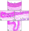
Figure 1
Figure 1
Photomicrographs of sections in the carotid artery stained with H&E. (A): In the control groups, the wall of the carotid artery consisted of thin tunica intima lined with endothelial cells (arrows), the intima mingles with the underlying media, which is formed from smooth muscle fibers (S), and wavy elastic fibers (corner arrow). The adventitia (A) is composed of loose connective tissue. (B,C): Reserpine-treated group. (B): Loss of some endothelial cells (crossed arrow) and the shedding of others (double arrowheads) in the lumen. Tunica media contains foam cells (arrowhead) and smooth muscle fibers with plump nuclei (zigzag arrows) or dark pyknotic nuclei (thick arrow). (C): Tunica media shows cytoplasmic vacuolation in the smooth muscle fibers (curved arrows). Tunica adventitia appears thick with inflammatory cell infiltration (double arrow) and mast cells (tailed arrows). (D): The fisetin-treated group demonstrated marked improvement of the wall and endothelial lining (arrows), apart from the loss of few epithelial cells (crossed arrow).
Figure 2
Figure 2
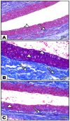
Photomicrographs of sections in the carotid artery stained with Masson’s trichrome stain. (A): In the control groups, minimal collagen fibers in tunica media (arrow) and in the tunica adventitia (arrowhead) are observed. (B): In the reserpine-treated group, a massive deposition of collagen fibers in tunica media (arrows) and in the tunica adventitia (arrowheads) is observed. (C): In the fisetin-treated group, moderate amounts of collagen fibers in tunica media (arrow) and in the tunica adventitia are observed(arrowhead).
Figure 3
Figure 3
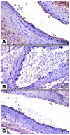
Photomicrographs of sections in the carotid artery immunohistochemically stained with eNOS. (A): In the control groups, strong positive eNOS immunoreactivity is observed in the endothelial lining of the tunica intima (arrowheads). (B): In the reserpine-treated group, decreased eNOS immunoreactivity (arrowheads) is observed (C): In the fisetin-treated group, weak positive eNOS immunoreactivity (arrowheads) in the endothelial lining of the tunica intima is observed.
Figure 4
Figure 4
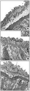
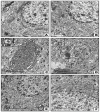
Electron micrographs of sections in the carotid arteries (A): In the control groups, the endothelial cells have a euchromatic nucleus (N), and its cytoplasm contains mitochondria (M) and few lysosomes (L). (B): In the reserpine-treated group, small heterochromatic nuclei (n) in the endothelial cells, mitochondria (m) with destructed cristae, numerous lysosomes (L), and autophagosome (A) are observed in the cytoplasm. Note the presence of apoptotic smooth muscle cells (asterisks) with a heterochromatic nucleus and condensed cytoplasm in the tunica media. The boxes indicate the destructed cristae of the mitochondria. (C): In the fisetin-treated group, normal endothelial cells with a euchromatic nucleus (N), normal mitochondria (M), and few lysosomes (L) are observed.
Figure 5
Figure 5
Photomicrographs of sections in the cerebral cortex stained with H& E. (A): In the control groups, large pyramidal cells and scattered granular ones can be observed. The pyramidal cells (P) have long apical dendrites, basophilic cytoplasm, and large vesicular nuclei. The granular cells (G) have rounded cell bodies with large rounded vesicular nuclei. The neuropil (white asterisk) contains the nuclei of neuroglial cells (thick arrow) and normal blood vessels (BV). (B,C): In the reserpine-treated group, (B) apoptotic cells (curved arrows) with small darkly stained nuclei and little acidophilic cytoplasm with vacuolation (black asterisk) in the neuropil are observed. (C): Perineuronal empty spaces (arrows) around deformed and shrunken pyramidal cells are visible. Vacuolation (black asterisk), inflammatory cellular infiltration (arrowhead), and dilated congested blood vessels (crossed arrow) are noticed in the neuropil. (D): In the fisetin-treated group, normal pyramidal (P) and granular (G) cells are seen. Few shrunken cells (arrows) with dark nuclei and perineuronal empty spaces and few apoptotic cells (curved arrow) are noticed. Normal neuropil (white asterisk) with non-congested blood vessels (BV) is seen.
Figure 6
Figure 6
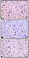
Photomicrographs of sections in the cerebral cortex immunohistochemically stained with eNOS. (A): In the control group, strong positive eNOS immunoreactivity is seen in the endothelial lining of the blood vessels (arrows) (B): In the reserpine-treated group, week positive eNOS immunoreactivity (arrows) can be observed (C): In the fisetin-treated group, mild positive eNOS immunoreactivity in the endothelial lining of blood vessels (arrows) can be seen.
Figure 7
Figure 7
Electron micrographs of sections in the cerebral cortex. (A,B): In the control groups, (A) pyramidal cells with long apical dendrites (AP) and rounded euchromatic nuclei (N) are observed. The cytoplasm shows rER cisternae (R), polyribosomes (arrows), few lysosomes (L), mitochondria (M) with normal cristae pattern, and Golgi saccules (G). Notice the presence of myelinated nerve fibers with the regular smooth contour of its myelin sheath (arrowhead) in the surrounding neuropil (B): A granular cell with a rounded euchromatic nucleus (N) and contains rER cisternae (R), polyribosomes (arrows), few lysosomes (L), and mitochondria (M) with a normal cristae pattern in the cytoplasm. (C,D): In the reserpine-treated group, (C) pyramidal cells show irregular heterochromatic nuclei (n) with corrugated nuclear membranes surrounded by a wide perinuclear space (zigzag arrows). Mitochondria with destructed cristae (m) autophagosome (A) and markedly dilated rER cisternae (crossed arrows) are seen in the cytoplasm. The nerve fibers (double arrowheads) in the surrounding neuropil have a disrupted myelin sheath. (D): The granular cell shows an irregular nucleus (n) with a corrugated nuclear membrane (zigzag arrows). The cytoplasm reveals dilated rER cisternae (crossed arrows), numerous lysosomes (L), and mitochondria (m) with destructed cristae. The boxes indicate the destructed cristae of mitochondria. (E,F): In the fisetin-treated group, (E) normal pyramidal cell with apical dendrites (AP) are observed. (F): Normal granular cell. In both figures (E,F), the nucleus (N) is rounded and euchromatic, and the cytoplasm contains average rER (R), mitochondria (M) with a normal cristae pattern, polyribosomes (arrows), and few lysosomes (L). Few mitochondria (m) with destructed cristae are noticed.

Similar articles

Cited by

References

    1. Jones G.T., Atzeni F., Beasley M., Flu B.E., Sarzi-Puttini P., Macfarlane G.J. The prevalence of fibromyalgia in the general population: A comparison of the American College of Rheumatology 1990, 2010, and modified 2010 classification criteria. Arthritis Rheumatol. 2015;67:568–575. doi: 10.1002/art.38905. - DOI - PubMed
    1. Choi D.H., Kim H.S. Quantitative analysis of nailfold capillary morphology in patients with fibromyalgia. Korean J. Intern. Med. 2015;30:531–537. doi: 10.3904/kjim.2015.30.4.531. - DOI - PMC - PubMed
    1. Son C.N., Kim S.H., Chang H.W., Kim J.M. A neurometabolite study of chronic daily headache in patients with systemic lupus erythematosus using magnetic resonance spectroscopy: Comparison with fibromyalgia patients and healthy controls. Korean J. Intern. Med. 2016;31:1171–1177. doi: 10.3904/kjim.2015.196. - DOI - PMC - PubMed
    1. Bradley L.A. Pathophysiology of fibromyalgia. Am. J. Med. 2009;122:S22–S30. doi: 10.1016/j.amjmed.2009.09.008. - DOI - PMC - PubMed
    1. Scherbakov N., Szklarski M., Hartwig J., Sotzny F., Lorenz S., Meyer A., Grabowski P., Doehner W., Scheibenbogen C. Peripheral endothelial dysfunction in myalgic encephalomyelitis/chronic fatigue syndrome. ESC Heart Fail. 2020;7:1064–1071. doi: 10.1002/ehf2.12633. - DOI - PMC - PubMed

MeSH terms