. 2021 Dec 24;11(1):55.
doi: 10.3390/cells11010055.

Bone Marrow Harbors a Unique Population of Dendritic Cells with the Potential to Boost Neutrophil Formation upon Exposure to Fungal Antigen

Affiliations

Bone Marrow Harbors a Unique Population of Dendritic Cells with the Potential to Boost Neutrophil Formation upon Exposure to Fungal Antigen

Marieke Goedhart et al. Cells. .

Abstract

Apart from controlling hematopoiesis, the bone marrow (BM) also serves as a secondary lymphoid organ, as it can induce naïve T cell priming by resident dendritic cells (DC). When analyzing DCs in murine BM, we uncovered that they are localized around sinusoids, can (cross)-present antigens, become activated upon intravenous LPS-injection, and for the most part belong to the cDC2 subtype which is associated with Th2/Th17 immunity. Gene-expression profiling revealed that BM-resident DCs are enriched for several c-type lectins, including Dectin-1, which can bind beta-glucans expressed on fungi and yeast. Indeed, DCs in BM were much more efficient in phagocytosis of both yeast-derived zymosan-particles and Aspergillus conidiae than their splenic counterparts, which was highly dependent on Dectin-1. DCs in human BM could also phagocytose zymosan, which was dependent on β1-integrins. Moreover, zymosan-stimulated BM-resident DCs enhanced the differentiation of hematopoietic stem and progenitor cells towards neutrophils, while also boosting the maintenance of these progenitors. Our findings signify an important role for BM DCs as translators between infection and hematopoiesis, particularly in anti-fungal immunity. The ability of BM-resident DCs to boost neutrophil formation is relevant from a clinical perspective and contributes to our understanding of the increased susceptibility for fungal infections following BM damage.

Keywords: bone marrow; dectin-1; dendritic cells; fungal infection; granulopoiesis; hematopoiesis; zymosan.


Conflict of interest statement

The authors declare no conflict of interest.

Figures
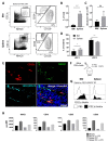
Figure 1
Figure 1
BM harbors a substantial population of DCs with an inflammatory profile. (A) Representative FACS plots of DCs and DC subsets in BM and spleen. (B,D) Percentages and (C) total numbers of DCs and DC subsets in BM and spleen. (Mean ± SD, n = 5–7, paired t-tests). *Total number of DCs in all BM containing bones were calculated per mouse by multiplying total number of BM DC derived from 2 femurs and 2 tibiae with the factor 3. (E) Cryosections of murine femurs stained for CD11c (red), MHCII (green), VE-cadherin and CD31 (cyan) and counterstained with Hoechst to visualize nuclei (blue). Original magnification = 630x. Scale bar = 50 µm. Representative image of 3 mice. (F) Experimental set-up for (G) in vivo binding of intravenously injected biotinylated CD11c to BM and splenic DCs within a 2-min time period. FACS plots are representative of 3 mice. (H) Quantification of geoMFI for expression of MHCII, CD40, CD80, and CD86 by DC from BM and spleen, in steady state, and 6 h after intravenous administration of LPS. (Mean ± SD, n = 2). * p < 0.05; ** p < 0.01; *** p < 0.001.
Figure 2
Figure 2
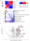
mRNA profiling reveals high expression of c-type lectin family receptors by BM DCs. RNAseq analysis was performed on cDC2 derived from BM and spleen. (A) Hierarchical clustering of the samples based on Pearson’s correlation coefficient. (B) Bar plot showing a total of 3697 differentially expressed (DE) genes being significantly up- (red, 2052) or downregulated (blue, 1645) in BM cDC2 as compared to splenic cDC2. (C) Hierarchical clustering of gene ontology (GO)-terms enriched in the DE expressed genes. In total 51 GO-terms were found enriched, which we clustered into 8 groups by comparison of their overlap index. Results are shown as a heatmap of the overlap indices, with the clustering and a color representation of each group on the left and a short description of the group on the right. The full heatmap is shown in Supplementary Figure S2. (D) Volcano plot of significantly up- and downregulated genes. Colored dots indicate genes encoding CTL/CTLD superfamily receptors being significantly up- (red) and downregulated (blue) in BM cDC2 as compared to splenic cDC2.
Figure 3
Figure 3
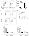
BM DCs express high, functional protein levels of the c-type lectin receptor Dectin-1. (A) Representative FACS plots and (B) quantification of geoMFI for expression of Dectin-1 by subsets in BM and spleen, as determined by flow cytometry (Mean ± SD, n = 2). (C) Representative FACS plots and (D) quantification of zymosan uptake by BM DCs and splenic DCs after 2 h in vitro co-culture (n = 3, p < 0.0001, 2-way ANOVA). (E) Uptake of zymosan particles by BM DCs and splenic DCs after 2 h in vitro co-culture in the presence or absence of a Dectin-1 blocking antibody (Mean ± SD, n = 3, multiple t-tests). (F) Uptake of life Aspergillus conidia by BM DCs and splenic DCs after overnight in vitro co-culture in the presence or absence of a Dectin-1 blocking antibody (Mean ± SD, n=3, multiple t-tests). * p < 0.05; *** p < 0.001.
Figure 4
Figure 4
Human BM DCs take up zymosan in a CR3-dependent manner. (A) Representative FACS plots of DCs in human BM aspirates. (Lineage: CD3, CD19, CD20, CD14, CD16, CD56). (B) Representative histograms of geoMFI for expression of Dectin-1, Dectin-2, CD11b, and CD18 expression by human BM DCs. Representative FACS plots (C) and quantification of uptake of zymosan by human BM DCs after 2 h (D) and overnight (E) in vitro co-culture in the presence or absence of CR3 blocking antibodies (Mean ± SD, n = 3, paired t-test). * p < 0.05.
Figure 5
Figure 5
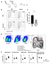
Zymosan is taken up by BM DCs after in vivo administration and boosts the number of neutrophil progenitors. (A) Experimental set-up, (B) representative FACS plots, and (C) quantification of in vivo uptake of zymosan particles by BM DCs and splenic DCs, 16 h after intravenous administration of zymosan. (Mean ± SD, n = 5, paired t-test). (D) Gating strategy for neutrophil precursors. (E) Quantification of neutrophil precursors in murine BM, 16 h after intravenous administration of zymosan. (Mean ± SD, n = 5 per group, unpaired t-test). * p < 0.05; ** p < 0.01; *** p < 0.001.
Figure 6
Figure 6
BM DCs promote neutrophil formation in a G-CSF dependent manner upon exposure to zymosan. (A) Experimental set-up of in vitro LSK cell cultures (B) Total number of cells, (C) percentage of LSK, and (D) percentage of CD11b+GR-1+ neutrophil type cells after 11 days in vitro culture of LSK cells with supernatant of BM DCs and zymosan cocultures. Graphs depict mean ± SD of experimental duplicates and are representative of 4 independent experiments. (unpaired t-test). Gating strategy for LSK cells and neutrophil type cells after culture can be found in Supplementary Figure S6. Graphs depict mean ± SD of experimental duplicates and are representative of three independent experiments. (unpaired t-test). ** p < 0.01.

Similar articles

Cited by

References

    1. Mazo I.B., Honczarenko M., Leung H., Cavanagh L.L., Bonasio R., Weninger W., Engelke K., Xia L., McEver R.P., Koni P.A., et al. Bone marrow is a major reservoir and site of recruitment for central memory CD8+ T cells. Immunity. 2005;22:259–270. doi: 10.1016/j.immuni.2005.01.008. - DOI - PubMed
    1. Slamanig S.A., Nolte M.A. The Bone Marrow as Sanctuary for Plasma Cells and Memory T-Cells: Implications for Adaptive Immunity and Vaccinology. Cells. 2021;10:1508. doi: 10.3390/cells10061508. - DOI - PMC - PubMed
    1. Milo I., Sapoznikov A., Kalchenko V., Tal O., Krauthgamer R., Van Rooijen N., Dudziak D., Jung S., Shakhar G. Dynamic imaging reveals promiscuous crosspresentation of blood-borne antigens to naïve CD8+ T cells in the bone marrow. Blood. 2013;122:193–208. doi: 10.1182/blood-2012-01-401265. - DOI - PubMed
    1. Feuerer M., Beckhove P., Garbi N., Mahnke Y., Limmer A., Hommel M., Hämmerling G.J., Kyewski B., Hamann A., Umansky V., et al. Bone marrow as a priming site for T-cell responses to blood-borne antigen. Nat. Med. 2003;9:1151–1157. doi: 10.1038/nm914. - DOI - PubMed
    1. Sapoznikov A., Pewzner-Jung Y., Kalchenko V., Krauthgamer R., Shachar I., Jung S. Perivascular clusters of dendritic cells provide critical survival signals to B cells in bone marrow niches. Nat. Immunol. 2008;9:388–395. doi: 10.1038/ni1571. - DOI - PubMed

Publication types

MeSH terms