. 2021 Dec 30;11(1):117.
doi: 10.3390/cells11010117.

MFUM-BrTNBC-1, a Newly Established Patient-Derived Triple-Negative Breast Cancer Cell Line: Molecular Characterisation, Genetic Stability, and Comprehensive Comparison with Commercial Breast Cancer Cell Lines

Affiliations

MFUM-BrTNBC-1, a Newly Established Patient-Derived Triple-Negative Breast Cancer Cell Line: Molecular Characterisation, Genetic Stability, and Comprehensive Comparison with Commercial Breast Cancer Cell Lines

Kristijan Skok et al. Cells. .

Abstract

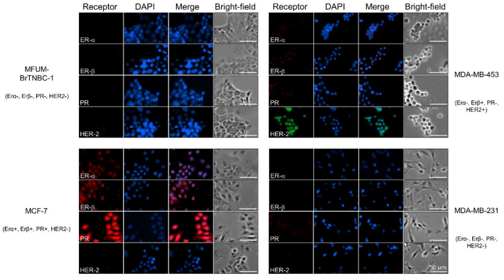
Triple-negative breast cancer (TNBC) is a breast cancer (BC) subtype that accounts for approximately 15-20% of all BC cases. Cancer cell lines (CLs) provide an efficient way to model the disease. We have recently isolated a patient-derived triple-negative BC CL MFUM-BrTNBC-1 and performed a detailed morphological and molecular characterisation and a comprehensive comparison with three commercial BC CLs (MCF-7, MDA-MB-231, MDA-MB-453). Light and fluorescence microscopy were used for morphological studies; immunocytochemical staining for hormone receptor, p53 and Ki67 status; RNA sequencing, qRT-PCR and STR analysis for molecular characterisation; and biomedical image analysis for comparative phenotypical analysis. The patient tissue-derived MFUM-BrTNBC-1 maintained the primary triple-negative receptor status. STR analysis showed a stable and unique STR profile up to the 6th passage. MFUM-BrTNBC-1 expressed EMT transition markers and displayed changes in several cancer-related pathways (MAPK, Wnt and PI3K signalling; nucleotide excision repair; and SWI/SNF chromatin remodelling). Morphologically, MFUM-BrTNBC-1 differed from the commercial TNBC CL MDA-MB-231. The advantages of MFUM-BrTNBC-1 are its isolation from a primary tumour, rather than a metastatic site; good growth characteristics; phenotype identical to primary tissue; complete records of origin; a unique identifier; complete, unique STR profile; quantifiable morphological properties; and genetic stability up to (at least) the 6th passage.

Keywords: MCF-7; MDA-MB-231; MDA-MB-453; MFUM-BrTNBC-1; breast cancer cell lines; hormonal receptors; triple-negative breast cancer.


Conflict of interest statement

The authors declare no conflict of interest.

Figures

Figure 1
Figure 1
Work protocol of comparative CL analysis. Legend: The picture depicts a stepwise protocol for establishing and characterising MFUM-BrTNBC-1 in adherence to international guidelines and standards. The steps include primary tissue derivation, subsequent isolation, culturing, comparative characterisation and data analysis. ER—oestrogen receptor, PR—progesterone receptor, TNBC—triple-negative breast cancer, STR—short tandem repeat, ICC—immunocytochemistry, RT-qPCR—reverse transcription quantitative real-time PCR.
Figure 2
Figure 2
Morphological characteristics of MFUM-BrTNBC-1 CL. Legend: (A) The primary MFUM-BrTNBC-1 culture; 4th passage (upper), 5th passage (lower). Images were taken at ×50 magnification on Zeiss Axiovert 40 inverted microscope. Scale bar = 200 μm (B) Immunofluorescence images of actin filaments (red) and nuclei (blue) showing cell morphology of MFUM-BrTNBC-1, MCF-7, MDA-MB-453 and MDA-MB-231 (cells were stained by using fluorescent phalloidin conjugate for actin and DAPI for nuclei). Immunofluorescence staining was carried out in triplicate for all CLs, and representative images are shown. Images were taken at ×10 magnification on EVOS FL fluorescence microscope). Scale bar = 50 μm.
Figure 3
Figure 3
Comparative characterisation of receptor status for human breast cancer cell lines MFUM-BrTNBC-1, MCF-7, MDA-MB-453 and MDA-MB-231. Legend: Immunofluorescence staining was carried out in triplicate for all CLs, and representative images are shown. ER-α—oestrogen α receptor, ER-β—oestrogen β receptor, PR—progesterone receptor, HER-2—human epidermal growth factor receptor 2. Scale bar = 50 mm.
Figure 4
Figure 4
Relative levels of hormone receptor transcripts (ER-α, PR and HER-2) by qRT-PCR. Legend: Relative levels of hormone receptor transcripts in MFUM-BrTNBC-1 and commercial BC CLs MCF-7, MDA-MB-453 and MDA-MB-231 were analysed by qRT-PCR. The level of ER and PR transcripts in MCF-7 cells is presented as 1, while the transcript level of HER-2 is presented as 1 in MDA-MB-453 cells. Comparison of receptor expression in MCF-7 (ER+ PR+ HER-2) versus other lines: * p < 0.05, Mann-Whitney U-test. Comparison of HER-2 expression in MDA-MB-453 (ER PR HER-2+) versus other lines: # p < 0.05, Mann–Whitney U-test. Differences in HER-2 expression between MFUM-BrTNBC-1 passages (MFUM 3rd, 4th, 5th and 6th p.): n.s. (not significant), ANOVA with Tukey post hoc test. Mean ± s.d. is shown.
Figure 5
Figure 5
Immunofluorescence staining of p53 and Ki67 in human breast cancer cell lines MFUM-BrTNBC-1, MCF-7, MDA-MB-453 and MDA-MB-231. Legend: Breast cancer cell lines were stained for either p53 (green) or Ki67 (green) and counterstained with DAPI (blue) to visualise the nuclei. The staining was carried out in triplicate for all cell lines and representative images are shown. Scale bar = 50 mm.
Figure 6
Figure 6
Expression of epithelial–mesenchymal transition (EMT) markers in MFUM-BrTNBC-1, MDA-MB-453 and MDA-MB-231 compared to MCF-7.

References

    1. Sung H., Ferlay J., Siegel R.L., Laversanne M., Soerjomataram I., Jemal A., Bray F. Global cancer statistics 2020: GLOBOCAN estimates of incidence and mortality worldwide for 36 cancers in 185 countries. CA. Cancer J. Clin. 2021;n/a doi: 10.3322/caac.21660. - DOI - PubMed
    1. Waks A.G., Winer E.P. Breast Cancer Treatment: A Review. JAMA - J. Am. Med. Assoc. 2019;321:288–300. doi: 10.1001/jama.2018.19323. - DOI - PubMed
    1. Milioli H.H., Tishchenko I., Riveros C., Berretta R., Moscato P. Basal-like breast cancer: Molecular profiles, clinical features and survival outcomes. BMC Med. Genomics. 2017;10:19. doi: 10.1186/s12920-017-0250-9. - DOI - PMC - PubMed
    1. The WHO Classification of Tumours Editorial Board, editor. Breast Tumors. 5th ed. International Agency for Research on Cancer; Lyon, France: 2019.
    1. Núñez Abad M., Calabuig-Fariñas S., Lobo de Mena M., José Godes Sanz de Bremond M., García González C., Torres Martínez S., García-García J.Á., Iranzo González-Cruz V., Camps Herrero C. Update on systemic treatment in early triple negative breast cancer. Ther. Adv. Med. Oncol. 2021;13:1758835920986749. doi: 10.1177/1758835920986749. - DOI - PMC - PubMed

Publication types