Review
. 2021 Dec 30;11(1):120.
doi: 10.3390/cells11010120.

Interleukin-8 in Melanoma Pathogenesis, Prognosis and Therapy-An Integrated View into Other Neoplasms and Chemokine Networks

Affiliations
Review

Interleukin-8 in Melanoma Pathogenesis, Prognosis and Therapy-An Integrated View into Other Neoplasms and Chemokine Networks

Anca Filimon et al. Cells. .

Abstract

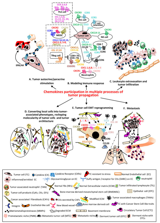
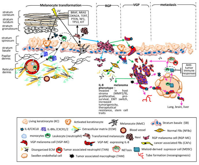
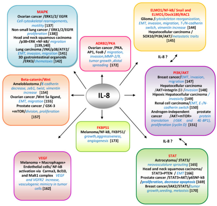
Cutaneous melanoma accounts for only about 7% of skin cancers but is causing almost 90% of deaths. Melanoma cells have a distinct repertoire of mutations from other cancers, a high plasticity and degree of mimicry toward vascular phenotype, stemness markers, versatility in evading and suppress host immune control. They exert a significant influence on immune, endothelial and various stromal cells which form tumor microenvironment. The metastatic stage, the leading cause of mortality in this neoplasm, is the outcome of a complex, still poorly understood, cross-talk between tumor and other cell phenotypes. There is accumulating evidence that Interleukin-8 (IL-8) is emblematic for advanced melanomas. This work aimed to present an updated status of IL-8 in melanoma tumor cellular complexity, through a comprehensive analysis including data from other chemokines and neoplasms. The multiple processes and mechanisms surveyed here demonstrate that IL-8 operates following orchestrated programs within signaling webs in melanoma, stromal and vascular cells. Importantly, the yet unknown molecularity regulating IL-8 impact on cells of the immune system could be exploited to overturn tumor fate. The molecular and cellular targets of IL-8 should be brought into the attention of even more intense scientific exploration and valorization in the therapeutical management of melanoma.

Keywords: CXCL8; CXCR1; CXCR2; chemokines; chemokines in tumor progression; interleukin-8.


Conflict of interest statement

The authors declare no conflict of interest.

Figures

Figure 1
Figure 1
The main processes regulated in various cellular phenotypes by CKs in tandem with their receptors. (A) TCssynthetize and secrete CKs which bind to self CKRs or CKRs expressed by other cells resulting activation of signaling pathways which sustain proliferation, survival and stress-resistance of the same TC (autocrine) or adjacent phenotypes (paracrine) [5]. (B) The CKs secreted by TCs recruit different immune cells in tumor micro environment (TME). The most relevant and documented examples of CKs/CKRs axes are presented. The pro-tumoral associated with poor prognosis are shown with dotted lines (NK—natural killer; Th1/Th2-CD4+T lymphocytes helper type1/type2; CD8+/CD4+-TIL-CD4+/CD8+ Tumor infiltrating lymphocyte; Treg-Regulatory T cell; MDSC—myeloid derived suppressor cell; DC—dendritic cell) [6,7]. (C) CKs secreted by TCs bound to the receptors (glycosaminoglycans and Duffy antigen) of inflamed (wrinkled) ECs are transcytosed and accumulated onto luminal face of ECs; the leucocytes migrate into lumen of the vessel toward CKs and establish firm attachments via overexpressed integrins, traverse endothelium through a gap junction or a pore, follow the extracellular CK gradient and enter the TME becoming TANs [8]. (D) Primary tumors contain NFBs and ECM with an ordered composition adequate to ensure immune infiltrates (left). Secreted factors by TCs convert NFBs and other Stromal Cells (SCs) into CAFs or Tumor Stromal Cells (TSCs-BMDMSCs or EPCs); here for simplicity only CAFs are presented. TSCs release various CKs which increase the number of stationary TAMs, that secrete CKs pro-tumor cell survival, induce angiogenesis, sustain the proliferative, migratory and invasive processes in TCs and vascular mimicry phenomenon, represented by Endothelial-like cells. The advanced tumor ECM composition becomes stiffer, includes modified proteins, stronger interactions via overexpressed integrins, keeping chemotherapeutic drugs and immune cells at tumor periphery (right) [9,10,11]. (E) The process of endothelial to mesenchymal transition (EMT) is essential in enabling TCs for invasion and metastatic dissemination. The most significant CKs secreted by TSCs acting on CKRs expressed on TCs and involved in EMT are presented [12]. (F) The cell signaling pathways activated by CKs in TCs and TSCs define the metastasizing process with the following major events: Inducing Cancer Stem Cell-like traits in TCs (self-renewal, colonies formation, mesenchymal multipotent differentiation) [13]. Remodeling ECM and invasion—the increased MMPs in TME disassemble components of ECM; TCs acquire invadopodia, protrude into the basement membranes, advance and disseminate within surrounding tissues [14]. Intravasation—TCs migrate across the vessel into blood or lymph circulatory systems and become CTCs [15]. Death or survival within circulatory system—until the opportunity of extravasation occurs, CTCs have to overcome hurdles as: lack of GFs/CytKs, the circulatory flow, anoikis, anti-tumor mechanisms of activated NKs/phagocytic cells and form circulating tumor microemboli [16]. Pre-metastatic niches (PMNs) formation—in secondary organs, as result of migration of hematopoietic bone marrow cells and SCs to particular sites in distal organs, with remodeled ECM, occur environmental milieus favorable to later installation and proliferation of TCs. These events facilitate the colonization with TCs, CyKs, GFs and TCs-derived exosomes as well as the suppression of immune system and installation of hypoxia. Distinct PMNs are formed in different organs [17,18]. In addition, dormant niches (DNs) (dotted line) with opposite properties from PMNs are also formed. Extravasation—TCs leave circulatory system and are seeded in PMNs [19]. Dissemination—Unlike DNs which will promote tumor cell dormancy, PMNs will sustain metastatic outgrowth instead. A primary tumor has the ability to seed more than one organ. PMNs are formed in different organs, which control the fate of TCs seeding at each metastatic site. A tumor contains sub-clones which colonize specific organs forming organ-specific metastasis [18].
Figure 2
Figure 2
IL-8 contribution to melanoma progression. Melanocyte transformation (black solid arrows)—in KCs the UV light induces secretion of various CKs including IL-8 which trigger extravasation of monocytes and neutrophils into dermis and epidermis (black dotted arrows) creating a pro-inflammatory site (dotted circle) [124,125]. In MelCs UV increases both protective melanin synthesis and ROS generation, destructively affecting MelC genome stability; the resulting transformed cells have mutations (the solid line box), are highly proliferative forming benign tumors (nevi). Neither MelCs nor nevus cells express endogenously IL-8. Radial growth phase (RGP) (blue arrows)—transformed cells proliferate along SB, forming a patch in the skin. The RGP-MCs invade dermis; however, they cannot form a tumor mass. The RGP-stage is generally associated with benignity and RGP-specimens still do not express IL-8. Vertical growth phase (VGP) and IL-8 phenotype (green arrows)—activated KCs together with hypoxia conditions and pro-inflammatory CyKs (IL-1 α, IL-1β, TNF-α) trigger IL-8 expression in RGP-MCs. They switch the molecular repertoire, activate processes which allow them to protrude beyond the SB and acquire IL-8 expression [103]. The MCs inhibitory motility signals are downregulated; MCs become invasive into papillary and reticular dermis, being actively involved in the transformation of NFBs into CAFs; CAFs increase melanoma invasiveness and ECM degradation. MCs expressing IL-8/IL-8Rs grow intensely due to autocrine/paracrine signals, attach to degraded ECM with overexpressed integrins and migrate deep into stromal compartment forming clusters (noduli) and in all these processes IL-8 is actively involved [104,105]. IL-8-VGP melanoma phenotype is correlated with an invasive potential, development of metastasis and poor prognosis. Metastasis (pink arrows)—MCs with IL-8/IL-8Rs encounter circulatory systems. IL-8 mediates paracrine mechanisms between MCs and neutrophils and MDSCs resulting their extravasation into tumor mass (dotted circle) [108]. TANs and NETs have positive or negative consequences on tumor fate [110] whereas MDSCs have intense suppressive anti-tumor immune activities [116]. In addition, TAMs secrete IL-8 and stimulate MCs to release IL-8 and other CyKs [113]. ECs altered because of pro-inflammatory milieu allow extravasation of immune cells, express IL-8Rs and exchange signals with MCs. The IL-8-mediated cross-talk between immune, tumor and ECs assist intravasation of MCs, their maintenance in circulation by adhering to ECs, further extravasation, colonization of secondary places (lung, brain, liver) and neovascularization and tube formation [122,123].
Figure 3
Figure 3
Molecular pathways IL-8-mediated and deciphered in various neoplasms.The main well-documented pathways and key-molecular actors involved in controlling IL-8-mediated processes tumor cell-specific are presented. The communication between different pathways as ELMO1/AKT/STAT possibly mediated by IL-8 are suggested.

References

    1. Payne A.S., Cornelius L.A. The Role of Chemokines in Melanoma Tumor Growth and Metastasis. J. Investig. Dermatol. 2002;118:915–922. doi: 10.1046/j.1523-1747.2002.01725.x. - DOI - PubMed
    1. Balkwill F. Cancer and the chemokine network. Nat. Rev. Cancer. 2004;4:540–550. doi: 10.1038/nrc1388. - DOI - PubMed
    1. Bonecchi R., Locati M., Mantovani A. Chemokines and Cancer: A Fatal Attraction. Cancer Cell. 2011;19:434–435. doi: 10.1016/j.ccr.2011.03.017. - DOI - PubMed
    1. Marcuzzi E., Angioni R., Molon B., Calì B. Chemokines and chemokine receptors: Orchestrating tumor metastasization. Int. J. Mol. Sci. 2019;20:96. doi: 10.3390/ijms20010096. - DOI - PMC - PubMed
    1. Wang J., Seethla R.R., Zhang Q., Gooding W., van Waes C., Hasegawa G., Ferris L.R. Autocrine and paracrine chemokine receptor 7 activation in head and neck cancer: Implications for therapy. J. Natl. Cancer Inst. 2008;100:502–512. doi: 10.1093/jnci/djn059. - DOI - PubMed

Publication types