. 2022 Jan 1;11(1):138.
doi: 10.3390/cells11010138.

Temporal Quantitative Phosphoproteomics Profiling of Interleukin-33 Signaling Network Reveals Unique Modulators of Monocyte Activation

Affiliations

Temporal Quantitative Phosphoproteomics Profiling of Interleukin-33 Signaling Network Reveals Unique Modulators of Monocyte Activation

Devasahayam Arokia Balaya Rex et al. Cells. .

Abstract

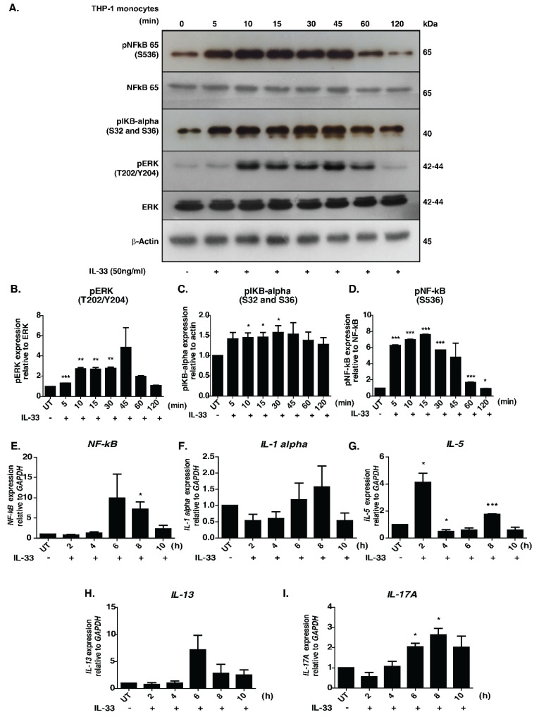
Interleukin-33 (IL-33), a member of the IL-1 superfamily cytokines, is an endogenous danger signal and a nuclear-associated cytokine. It is one of the essential mediators of both innate and adaptive immune responses. Aberrant IL-33 signaling has been demonstrated to play a defensive role against various infectious and inflammatory diseases. Although the signaling responses mediated by IL-33 have been previously reported, the temporal signaling dynamics are yet to be explored. To this end, we applied quantitative temporal phosphoproteomics analysis to elucidate pathways and proteins induced by IL-33 in THP-1 monocytes. Employing a TMT labeling-based quantitation and titanium dioxide (TiO2)-based phosphopeptide enrichment strategy followed by mass spectrometry analysis, we identified and quantified 9448 unique phosphopeptides corresponding to 3392 proteins that showed differential regulation. Of these, 171 protein kinases, 60 phosphatases and 178 transcription factors were regulated at different phases of IL-33 signaling. In addition to the confirmed activation of canonical signaling modules including MAPK, NFκB, PI3K/AKT modules, pathway analysis of the time-dependent phosphorylation dynamics revealed enrichment of several cellular processes, including leukocyte adhesion, response to reactive oxygen species, cell cycle checkpoints, DNA damage and repair pathways. The detailed quantitative phosphoproteomic map of IL-33 signaling will serve as a potentially useful resource to study its function in the context of inflammatory and pathological conditions.

Keywords: DNA damage; DNA repair; IL-33; cytokine signaling; inflammation; mass spectrometry; phosphoproteomics.


Conflict of interest statement

The authors declare no conflict of interest.

Figures

Figure 1
Figure 1
Effect of IL-33 treatment on the MAPK and NFκB signaling cascades in THP-1 monocytes. (A). Western blot analysis showing the effect of IL-33 (50 ng/mL) on the phosphorylation status of NF-kB-p65 (S536), IKBα (S32/36), total NFKB p65, total ERK1/2 and phosphorylated ERK1/2 (T202/Y204) at different time points (5, 10, 15, 30, 45, 60, and 120 min). Densitometry analysis of the Western blots for (B) ERK1/2, phosphorylated ERK 1/2, (C) phosphorylated IκBα, (D) NFκB-p65 and phosphorylated NFκB-p65. The relative fold changes are shown. * p < 0.05 compared to control (Mean ± SEM, n = 3). Further, IL-33 treatment induced mRNA expression of various cytokines, including (E) NF-kB, (F) IL-1alpha, (G) IL-5, (H) IL-13 and (I) IL-17A. Quantitative real-time PCR was carried out after stimulating THP-1 monocytes with IL-33(50 ng/mL for varying durations (2, 4, 6, 8 and 10 h). The results are shown as fold change with respect to control cells (0 min). All the experiments were repeated in triplicates. * p < 0.05 compared to control (Mean ± SEM, n = 3). ** and *** denote medium and highly significant values respectively.
Figure 2
Figure 2
Temporal phosphoproteomics and proteomic analysis of THP-1 monocytes stimulated with IL-33. (A) Brief workflow depicting quantitative proteomic analysis of IL-33 stimulated THP-1 monocytes. THP-1 cells were stimulated with recombinant human IL-33 for indicated time points. Proteins were extracted from the cell lysates and subjected to in-solution trypsin digestion, followed by TMT-based chemical tagging for quantitative proteome profiling. The samples were analyzed on a Thermo Scientific™ Orbitrap Fusion™ Tribrid™ Mass Spectrometer. Mascot and SEQUEST algorithms were used for database searches. (B) Summary of the IL-33-induced phosphoproteome and total proteome. (C) Gene ontology analysis depicting the enriched biological processes for the IL-33-induced hyperphosphorylation events (D) Kinase enrichment analysis identified predicted upstream kinases for the phosphoproteins activated by IL-33. (E) Graph representing the top eight phosphatase families that were identified as being regulated by IL-33.
Figure 3
Figure 3
A snapshot of IL-33 signaling in THP-1 monocytes. (A) Brief schematic of the IL-33 pathway showing proteins and phosphosites identified by the phosphoproteome analysis. Temporal changes in IL-33 pathway phosphopeptides of: (B) TAB2/3 and MAPK14 modules; (C) IKBK-NFKB1 modules; (D) MAPK and PI3K/AKT modules; and (E) transcription factors and interleukins.
Figure 4
Figure 4
(A) Reactome pathway analysis of upregulated phosphoproteins showed induction of several signaling pathways including DNA damage and DNA repair pathways (highlighted with dotted lines). Further detailed analysis identified specific phosphosite changes in several DNA damage/repair proteins including (B) TP53BP1, (C) BRCA1, (D) NDRG1, (E) NUCKS1, (F) UNG, (G) MSH6, (H) POLD3, (I) XRCC1, (J) PARP1 and (K) SIRT1.

References

    1. Shipman L. Allergy: Neonatal IL-33 drives allergy. Nat. Rev. Immunol. 2017;17:80–81. doi: 10.1038/nri.2016.149. - DOI - PubMed
    1. Oboki K., Ohno T., Kajiwara N., Arae K., Morita H., Ishii A., Nambu A., Abe T., Kiyonari H., Matsumoto K., et al. IL-33 is a crucial amplifier of innate rather than acquired immunity. Proc. Natl. Acad. Sci. USA. 2010;107:18581–18586. doi: 10.1073/pnas.1003059107. - DOI - PMC - PubMed
    1. Kamijo S., Takeda H., Tokura T., Suzuki M., Inui K., Hara M., Matsuda H., Matsuda A., Oboki K., Ohno T., et al. IL-33-mediated innate response and adaptive immune cells contribute to maximum responses of protease allergen-induced allergic airway inflammation. J. Immunol. 2013;190:4489–4499. doi: 10.4049/jimmunol.1201212. - DOI - PubMed
    1. Humphreys N.E., Xu D., Hepworth M.R., Liew F.Y., Grencis R.K. IL-33, a potent inducer of adaptive immunity to intestinal nematodes. J. Immunol. 2008;180:2443–2449. doi: 10.4049/jimmunol.180.4.2443. - DOI - PubMed
    1. Marzullo A., Ambrosi F., Inchingolo M., Manca F., Devito F., Angiletta D., Zito A., Scicchitano P., Ciccone M.M. ST2L Transmembrane Receptor Expression: An Immunochemical Study on Endarterectomy Samples. PLoS ONE. 2016;11:e0156315. doi: 10.1371/journal.pone.0156315. - DOI - PMC - PubMed

Publication types