. 2022 Jan 4;11(1):157.
doi: 10.3390/cells11010157.

Recognition and Chaperoning by Pex19, Followed by Trafficking and Membrane Insertion of the Peroxisome Proliferation Protein, Pex11

Affiliations

Recognition and Chaperoning by Pex19, Followed by Trafficking and Membrane Insertion of the Peroxisome Proliferation Protein, Pex11

Katarzyna M Zientara-Rytter et al. Cells. .

Abstract

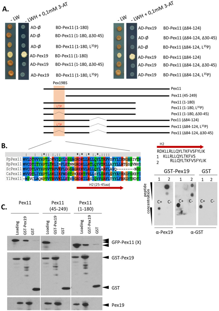
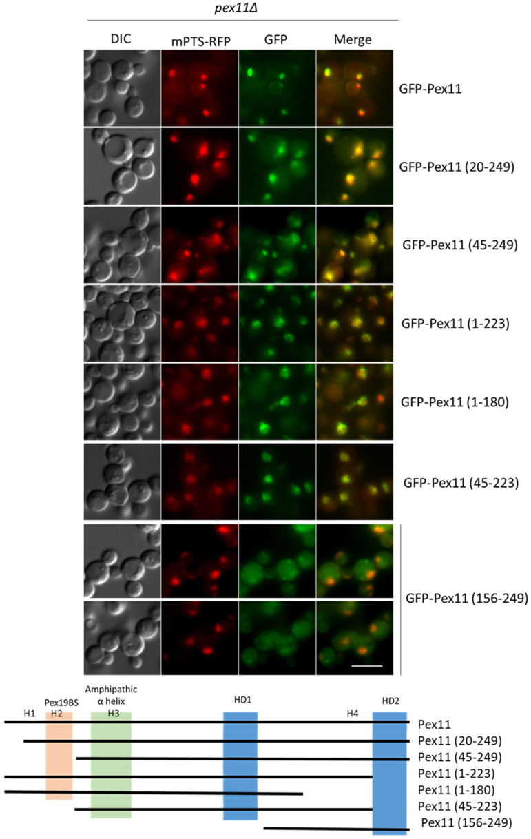
Pex11, an abundant peroxisomal membrane protein (PMP), is required for division of peroxisomes and is robustly imported to peroxisomal membranes. We present a comprehensive analysis of how the Pichia pastoris Pex11 is recognized and chaperoned by Pex19, targeted to peroxisome membranes and inserted therein. We demonstrate that Pex11 contains one Pex19-binding site (Pex19-BS) that is required for Pex11 insertion into peroxisomal membranes by Pex19, but is non-essential for peroxisomal trafficking. We provide extensive mutational analyses regarding the recognition of Pex19-BS in Pex11 by Pex19. Pex11 also has a second, Pex19-independent membrane peroxisome-targeting signal (mPTS) that is preserved among Pex11-family proteins and anchors the human HsPex11γ to the outer leaflet of the peroxisomal membrane. Thus, unlike most PMPs, Pex11 can use two mechanisms of transport to peroxisomes, where only one of them depends on its direct interaction with Pex19, but the other does not. However, Pex19 is necessary for membrane insertion of Pex11. We show that Pex11 can self-interact, using both homo- and/or heterotypic interactions involving its N-terminal helical domains. We demonstrate that Pex19 acts as a chaperone by interacting with the Pex19-BS in Pex11, thereby protecting Pex11 from spontaneous oligomerization that would otherwise cause its aggregation and subsequent degradation.

Keywords: Pex11; Pex19; peroxisomal membrane protein; peroxisome division; peroxisome proliferation protein.


Conflict of interest statement

The authors declare no conflict of interest.

Figures

Figure 1
Figure 1
Graphical representation of the Pex11 protein in yeast and humans. Graphical representation of the domain/motif architecture organization for each Pex11 protein in yeast S. cerevisiae, P. pastoris and in humans. Proteins are drawn to scale with the amino acids (aa) number indicated at the bottom. Most, but not all (e.g., HsPex11γ), Pex11-family proteins have four putative amphipathic helices, whose coordinates in PpPex11 are as follows—H1 (aa14-19), H2 (aa25-45), H3 (aa55-86), and H4 (aa203-218), shown as yellow, orange, green, and grey rectangles, respectively) and two hydrophobic domains (HD1 and HD2, blue rectangles).
Figure 2
Figure 2
PpPex11 contains one Pex19-binding site (Pex19-BS) near its N-terminal end. Pex11 interacts with Pex19 via its Pex19-BS. (A) Y2H screen to determine the location of the Pex19-BS within PpPex11. Two truncated forms of PpPex11 were subjected to site-directed mutagenesis within H2 region of Pex11 and fused with the BD domain of GAL4 to test their abilities to interact with AD-Pex19. Schematic representation of Pex11 mutants used in this study is shown and the H2 helix (aa25-45), within which the Pex19-BS was mapped for other yeast Pex11s, is highlighted in orange. 3-AT, 3-amino-1,2,4-triazole; AD, activation domain; BD, DNA binding domain; -LW, yeast synthetic drop-out medium without leucine and tryptophan serving as a positive control to show equal plating of cells; -LWH, yeast synthetic drop-out medium without leucine, tryptophan, and histidine. (B) Precise mapping of the Pex19-BS. Based on the location of the Pex19-BS in the H2 helix of Pex11, 14-mer (aa27-40) and 16-mer (aa30-45) peptides, with a 3aa shift between them, were synthesized, spotted onto a nitrocellulose membrane, and subjected to the in vitro Pex19 binding assay. Peptides and proper positive (C+) and negative (C) controls were spotted at two concentrations (20 nmol and 5 nmol, respectively) and tested for interaction with GST-PpPex19. Bound protein was detected immunologically with polyclonal anti-Pex19 or anti-GST antibodies. As a control, free GST and monoclonal anti-GST antibodies were used. Sequence alignment of N-terminal regions of Pex11 proteins from various species, showing conservation of specific residues within the H2 helix, is included. Residues in H2 helix are colored by Clustal X [38] based on their physico-chemical properties: hydrophilic, charged: D, E (magenta), K, R, H (red); hydrophilic, neutral: S, T, Q, N (green); hydrophobic: A, V, L, I, M, W, F (blue); P (yellow); G (orange; and other aromatic Y and H (cyan). Abbreviations and accessions numbers used in sequence alignments: Pp—Pichia pastoris, CAY69135; Hp—Hansenula polymorpha, DQ645582; Sc—Saccharomyces cerevisiae, CAA99168; Ca—Candida albicans, EAK92906; Yl—Yarrowia lipolytica, CAG81724. (C) The Pex11 (45-249) deletion mutant does not bind Pex19 in vitro. Binding studies revealed that removal of N-terminal end of Pex11 (aa1-44) is sufficient to inhibit Pex11–Pex19 binding in vitro. Only the mutant, Pex11 (45-249), was deficient in Pex19 binding, whereas the full-length and other deletion mutants, Pex11 (1-180), were still pulled down with GST-Pex19. Free GST protein was used as a control. There was equivalent loading in the input and bound lanes. Proteins were detected by anti-GFP, anti-GST, and anti-Pex19 antibodies.
Figure 3
Figure 3
The Pex19-BS of Pex11 binds to Pex19 via its hydrophobic surface and flanking positively-charged residues. Identification of H2 mutants of Pex11 deficient in Pex19 binding by dot-blot analysis. (A) The H2 peptides generated, including one harboring the HsPex11β H2 sequence but lacking Pex19-binding properties, as well as the PpPex11 H2 WT or mutant sequences (sequence details are indicated and mutated residues are marked in red) are shown on left. Dot-blot analysis (shown on right) was done as described in Figure 2B. Peptides were spotted on a nitrocellulose membrane at two concentrations (20 nmol and 5 nmol) and tested for interaction with GST-PpPex19. The top panel shows spotted peptides stained with Ponceau S prior to GST-PpPex19 addition. Bound protein was detected immunologically with polyclonal anti-Pex19 antibodies in the middle (short exposure) and bottom (long exposure) panels. (B) Helical-wheel diagrams of HsPex11β and PpPex11 H2 variants using HeliQuest. Positively-charged residues are shown in blue, negatively-charged residues in red, and hydrophobic residues in yellow. In addition, Ser and Thr are shown in purple, Gly and Ala in gray, Asn and Gln in pink, and His in sky blue. The arrows represent the helical hydrophobic moment. (C) CD spectra of the PpPex11 H2 wild-type and mutant peptides. The spectrum shows that H2 and its mutant variant H2 L8A, Y10A are unstructured in phosphate buffer, but the addition of 30% of TFE induces changes in the spectrum, typical for α-helical structures.
Figure 4
Figure 4
Pex11 is chaperoned by Pex19, which determines its stability via direct interaction with the Pex19-BS of Pex11. (A). Western blot analyses of GFP-Pex11 and other protein levels in WT and pex19Δ strains after methanol induction. WT and pex19Δ cells expressing Pex11-2HA from its endogenous PEX11 promoter, or the constitutive GAPDH promoter, were grown in methanol medium, and 2 OD cells were collected at the indicated time points. GFP-Pex11 levels were visualized by anti-GFP antibodies. Pex3 and Pex19 were detected using custom antibodies, and F1β was used as loading control. (B). Western blot analysis of Pex11-2HA and other protein levels at various time points in peroxisome-deficient strains after methanol induction. WT, pex19Δ, and pex3Δ cells in an atg30Δ (pexophagy-deficient) background expressing Pex11-2HA from its endogenous promoter were grown in methanol medium, and 2 OD cells were collected at indicated time points. Endogenous PMPs were detected with indicated antibodies, and F1β was used as loading control. SE—short exposure and LE—long exposure. (C). Removal of Pex19-BS destabilizes Pex11. Western blot analysis of levels of truncated forms of GFP-Pex11 at various time points in atg30Δ cells after methanol induction. GFP-Pex11, GFP-Pex11 (45-249), GFP-Pex11 (1-223) (referred to collectively as GFP-Pex11 (X)), and free GFP were expressed from the PEX11 promoter in the atg30Δ strain in methanol medium, and 2 OD cells were collected at indicated time points. Proteins were identified using respective antibodies. SE, short exposure and LE, long exposure.
Figure 5
Figure 5
N-terminal amphipathic helices H2 and H3 enable Pex11 dimerization. (A). Involvement of N-terminal helices of Pex11 in its self-interaction with Y2H. Full-length Pex11 and its truncated forms, as well as their mutated variants, were fused to either BD or AD domains of GAL4. As negative controls, empty pGBKT7 and pGADT7 were used. Different combinations of these vectors were transformed into the yeast strain Y2H Gold for mapping the regions involved in Pex11 dimerization. Schematic representation of Pex11 truncated and mutant forms used in this study. The H2 helix (aa25-45) containing the Pex19-BS is highlighted in orange, and the H3 (aa55-86) helix, known for lipid binding and dimerization properties, is in green. Sequence alignment of the N-terminal part of Pex11 proteins from various species, showing conservation of specific residues within the H3 helix, whose residues are colored as in Figure 1. Abbreviations and accessions numbers used in sequence alignments: Pp—Pichia pastoris, CAY69135; Hp—Hansenula polymorpha, DQ645582; Sc—Saccharomyces cerevisiae, CAA99168; Ca—Candida albicans, EAK92906; Yl—Yarrowia lipolytica, CAG81724. 3-AT, 3-amino-1,2,4-triazole; AD, activation domain; BD, DNA binding domain. (B). Pex11 preferentially dimerizes via its H2 helix. Pull-down assays using biotinylated peptides corresponding to the H2, H3, or H4 helices (sequences indicated in the figure) bound to streptavidin-coated resin as a bait and His6-GFP-Pex11 as a prey. Resins were washed, and proteins were eluted and analyzed by SDS-PAGE. His6-GFP-Pex11 was detected by immunoblotting with anti-GFP antibody, and the presence of peptides on the resin was verified by HRP-conjugated streptavidin. Shown is 25% of the input. SE, short exposure; ME, moderate exposure. Quantification of the pull-down assay is shown on the right, with the averages and standard deviations based on three independent sets of experiments. Western blot signals were quantified using the program ImageJ. (C). Same as panel B, except full-length His6-GFP-Pex11 or truncated form His6-GFP-Pex11 (45-249) was used as a prey. Quantification of the Western blots from three independent experiments is shown on the right with standard deviations. (D). Graphical representation of homotypic (H2-H2 or H3-H3) and heterotypic (H2-H3) interactions that drive Pex11 dimerization. The thickness of the arrows reflects the strength of the interactions detected by in vitro pull-down assays.
Figure 6
Figure 6
Pex19 binding and Pex11 dimerization to the H2 helix are mutually exclusive. (A). Dimerization of Pex11 via its H2 helix involves the same surface as that required for Pex19 binding. Identification of H2 mutants deficient in binding to Pex11 by in vitro pull-down. Biotinylated H2 peptides harboring either the HsPex11β H2 sequence (negative control) or Pex11 H2 WT (positive control) or mutant sequences (see Figure 2 for sequence details) were bound to streptavidin-coated resin prior to the addition of a 4-fold molar excess of His6-GFP-Pex11. Levels of captured His6-GFP-Pex11 were examined on SDS-PAGE and detected by immunoblotting with anti-GFP antibody. The presence of peptides on the resin was verified by HRP-conjugated streptavidin. Input was diluted 1:4 or 1:6 prior to loading. SE, short exposure; ME, moderate exposure. (B). Pex19 competes with Pex11 for the access to H2. Pull-down competition assays of the interaction between the H2 or H3 peptide and His6-GFP-Pex11 or His6-GFP-Pex11 (45-249) in competition with increasing amounts of the GST-Pex19 or GST alone used as a control are shown. GST-Pex19 or GST alone were added to the resin with bound H2 or H3 simultaneously with His6-GFP-Pex11 or His6-GFP-Pex11 (45-249). Resins were washed, and proteins were eluted and analyzed by SDS-PAGE. His6-GFP-Pex11s, GST-Pex19, and free GST were detected with respective antibodies, and presence of peptides on the resin was verified by HRP-conjugated streptavidin. A graphical illustration of possible interactions for each combination is included.
Figure 7
Figure 7
Pex11 import to peroxisomal membranes, but not its trafficking to peroxisomes, requires Pex19 binding. (A). Both full-length (GFP-Pex11) and truncated (GFP-Pex11 (45-249)) fusion proteins, respectively, colocalize with the peroxisomal membrane marker, mPTS-RFP. The pex11Δ cells containing mPTS-RFP and expressing GFP-tagged Pex11 variants from the endogenous PEX11 promoter were grown in methanol medium for 5 h (upper panel) or 16 h (o/n-lower panel) for fusion protein induction, prior to observation by fluorescence microscopy. Schematic representation of Pex11 forms used for live-cell imaging is shown below microscopy pictures. Positions of known and predicted modules are highlighted: Pex19-BS (orange), amphipathic helix (green), and hydrophobic regions (HD1 and HD2) predicted to be buried in lipid bilayers (blue). (B). Full-length Pex11, but not the Pex11 (45-249) truncated form, is an integral membrane protein. Western blot of membrane protein extraction assay in which the organelle membrane fraction from pex11Δ strains expressing Pex11-2HA, GFP-Pex11, and GFP-Pex11 (45-249) were resuspended in four different buffers for peripheral membrane protein extraction (Tris buffer pH 8, 2 mM Urea in Tris buffer pH 8, 0.1 M Na2CO3 pH 11.5, and Tris buffer pH 8 with Triton X-100) for 30 min at room temperature and fractionated by ultracentrifugation to obtain supernatant (S) and pellet (P) fractions. Proteins were visualized with anti-GFP, anti-HA, and Pex3 antibodies. Pex3 protein was used as a reference integral membrane protein. Note that for GFP-Pex11, the arrowhead indicates proper protein size, and asterisk is probably a truncated form. (C). Graphical representation of results. Model (A) presents Pex11 trafficking and incorporation into peroxisomal membranes with the assistance of Pex19 (and Pex3). Model (B) presents Pex11 (45-249) trafficking to, but not its insertion, into the peroxisome membrane, due to the lack of Pex19-BS. It is unclear if Pex11 (45-249) requires interaction with another protein (marked as “?”) for its targeting to peroxisomes. Bar = 5 μm.
Figure 8
Figure 8
Pex11 has a Pex19-independent mPTS that is distinct from the Pex19-BS. Fluorescence microscopy images of pex11Δ cells expressing full-length and truncated GFP-Pex11 fusion proteins and mPTS-RFP for peroxisome visualization after 5 h in methanol medium. Bar = 5μm. A schematic representation of Pex11 truncated forms used in this study is shown below the micrographs. Positions of known and predicted modules are highlighted as shown in Figure 5: Pex19-BS (orange); amphipathic helix (green); and hydrophobic regions, HD1, and HD2, predicted to be buried in lipid bilayers (blue).
Figure 9
Figure 9
Amphipathic helix H4 of Pex11 has a Pex19-independent mPTS. (A). Fluorescence microscopy images of pex11Δ cells expressing full-length and truncated GFP-Pex11 fusion proteins and mPTS-RFP for peroxisome visualization after 16 h in methanol medium. A schematic representation of Pex11 truncated forms used in this study is shown below the micrographs. Positions of known and predicted modules are highlighted similarly as in Figure 5. Bar = 5 μm. (B). The H4 (aa203-218) region in Pex11 is not recognized by Pex19. Short 14 and 15-mer peptides with three amino acid shifts spanning the identified mPTS with its flanking N- and C-terminal residues were synthesized and subjected to the Pex19 in vitro binding assay. Dot-blot analysis was done as described in Figure 2B. Peptides and proper controls (including peptides described in Figure 2B) were spotted on a nitrocellulose membrane at two concentrations (20 nmol and 5 nmol) and tested for interaction with GST-Pex19. Bound protein was detected immunologically with polyclonal anti-Pex19 antibodies. Sequence alignment of regions from various Pex11s corresponding to identified mPTS in Pex11 is shown at the bottom. Residues were colored by Clustal X [38], based on their physico-chemical properties as in Figure 2B. The position of the amphipathic helix H4 in Pex11 is marked by a red arrow, and its helical wheel plot generated using HeliQuest is shown on the right. The black arrow in the plot points to the hydrophobic face, and its length corresponds to the hydrophobic moment. (C). The secondary structures of the H3 (used as reference) and H4 peptides were analysed by CD spectroscopy. The spectrum shows that, like the H3 peptide, H4 is unstructured in phosphate buffer, but α-helical in 30% TFE. Abbreviations and accession numbers used in sequence alignments: Pp—Pichia pastoris, CAY69135; Hp—Hansenula polymorpha, DQ645582; Sc—Saccharomyces cerevisae, CAA99168; Ca—Candida albicans, EAK92906; Yl—Yarrowia lipolytica, CAG81724.

References

    1. Islinger M., Voelkl A., Fahimi H.D., Schrader M. The peroxisome: An update on mysteries 2.0. Histochem. Cell Biol. 2018;150:443–471. doi: 10.1007/s00418-018-1722-5. - DOI - PMC - PubMed
    1. Wanders R.J., Waterham H.R. Biochemistry of mammalian peroxisomes revisited. Annu. Rev. Biochem. 2006;75:295–332. doi: 10.1146/annurev.biochem.74.082803.133329. - DOI - PubMed
    1. Subramani S. Protein import into peroxisomes and biogenesis of the organelle. Annu. Rev. Cell Biol. 1993;9:445–478. doi: 10.1146/annurev.cb.09.110193.002305. - DOI - PubMed
    1. Motley A.M., Hettema E.H. Yeast peroxisomes multiply by growth and division. J. Cell Biol. 2007;178:399–410. doi: 10.1083/jcb.200702167. - DOI - PMC - PubMed
    1. Geuze H.J., Murk J.L., Stroobants A.K., Griffith J.M., Kleijmeer M.J., Koster A.J., Verkleij A.J., Distel B., Tabak H.F. Involvement of the endoplasmic reticulum in peroxisome formation. Mol. Biol. Cell. 2003;14:2900–2907. doi: 10.1091/mbc.e02-11-0734. - DOI - PMC - PubMed

Publication types