Review
. 2021 Dec 31;11(1):210.
doi: 10.3390/jcm11010210.

Surgery and Catheter Ablation for Atrial Fibrillation: History, Current Practice, and Future Directions

Affiliations
Review

Surgery and Catheter Ablation for Atrial Fibrillation: History, Current Practice, and Future Directions

Patrick M McCarthy et al. J Clin Med. .

Abstract

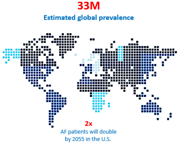
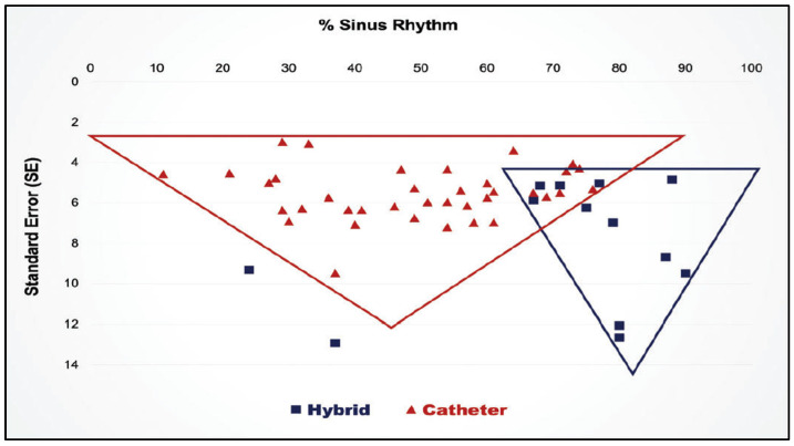
Atrial fibrillation (AF) is the most common of all cardiac arrhythmias, affecting roughly 1% of the general population in the Western world. The incidence of AF is predicted to double by 2050. Most patients with AF are treated with oral medications and only approximately 4% of AF patients are treated with interventional techniques, including catheter ablation and surgical ablation. The increasing prevalence and the morbidity/mortality associated with AF warrants a more aggressive approach to its treatment. It is the purpose of this invited editorial to describe the past, present, and anticipated future directions of the interventional therapy of AF, and to crystallize the problems that remain.

Keywords: atrial fibrillation; catheter ablation; electrophysiologic mapping; maze procedure.


Conflict of interest statement

McCarthy: Edwards Lifesciences: royalties and speaking fees; AtriCure and Medtronic: speaking fees. Cox: Consultant and Stockholder: AtriCure, Inc. and SentreHEART, Inc.; consultant, stockholder, and board of directors: Adagio Medical, Inc., PAVmed, Inc., Lucid Diagnostics, Inc. All other authors declare no conflict of interest.

Figures

Figure 1
Figure 1
Atrial fibrillation is an epidemic and the patient population with an estimated 33 million people suffering from the anomaly globally and that number is predicted to double in the next 15 years. Moreover, 30% of those patients could benefit from interventional therapy, but only 9% are treated with catheter ablation or surgery annually.
Figure 2
Figure 2
Trends in total numbers of surgical ablation procedures in the United States from 1 July 2011 through 30 June 2014. AVR = aortic valve replacement; CABG = coronary artery bypass graft surgery; MVRR = mitral valve repair or replacement (reproduced with permission from Badhwar et al.) [5].
Figure 3
Figure 3
The life expectancy following the first in-hospital diagnosis of atrial fibrillation in Medicare patient ranks eleventh if compared to the life expectancy following the diagnosis of the 25 most lethal cancers in the United States. The life expectancy is shorter following the diagnosis of atrial fibrillation than it is for such common neoplasms as colon cancer, melanoma, and breast cancer.
Figure 4
Figure 4
The impact of atrial fibrillation on the 20-year survival rate for women is much greater than the impact of atrial fibrillation in men (reproduced with permission from Stewart et al.) [10].
Figure 5
Figure 5
The experimental model created to map and treat atrial fibrillation in which mitral insufficiency is created without placing any lesions in the atrium and without causing any pericardial adhesions by violating the pericardial space.
Figure 6
Figure 6
A three-dimensional anatomic reconstruction of the atria using gated ECG-MRI scans with each of the MRI slices “stacked” on one another. This figure is taken from a movie that was created in our experimental laboratories in 1986.
Figure 7
Figure 7
Three-dimensional electrode arrays used for the experimental mapping (upper panel) and clinical mapping (lower panel) of atrial fibrillation in the mid-1980s. The individual bipolar electrodes were newly designed “target electrodes” that had the anode as the central point and the cathode in a circular pole surrounding the central anode. This design eliminated potential artifact cause by differences in the direction of wavefront propagation. The three-dimensional electrical data was then superimposed on the three-dimensional anatomic reconstruction of the same atria using gated MRI scans (Figure 6). The experimental electrode arrays contained 256 individual bipolar electrodes and the clinical electrode arrays contained 156 individual bipolar electrodes. Both electrode arrays were designed and created by Dr. John P. Boineau and Dr. Richard B. Schuessler.
Figure 8
Figure 8
Photograph of both atria immediately following the performance of a maze procedure in a canine experiment. The right coronary artery was injected with blue dye and the left coronary artery was injected with red dye to detect any evidence of devascularization of either atrium. There was none.
Figure 9
Figure 9
In order to document that atrial transport function persisted following an experimental canine maze procedure, an electromagnetic flow probe was placed around the ascending aorta. A-V sequential pacing was initiated and the aortic flow was recorded and remained stable for several minutes. The atrial pacing wire was then suddenly disconnected from the pacemaker, leaving the heart being paced in the ventricle only with no atrial contribution to forward aortic flow. The aortic flow immediately decreased by approximately 20% during ventricular pacing only. The atrial pacing wire was then reconnected to the pacemaker to resume A-V sequential pacing. The aortic flow immediately returned to its normal level, proving that the atrial contribution to forward aortic blood flow was approximately 20% following a maze procedure.
Figure 10
Figure 10
In 1997, the first minimally invasive surgical procedure for atrial fibrillation was performed. Because the far left side of the left atrium, which often had a bleeder from a branch of the circumflex coronary artery following a standard cut-and-sew maze procedure, could not be reached through the small mini-thoracotomy in the right fourth intercostal space, all of the lesions of the minimally invasive maze procedure had to be created with a cryoprobe rather than with a scalpel or scissors. This procedure was called the minimally invasive cryo-maze III procedure, and it has remained largely unchanged for the past 25 years (reproduced with permission from Cox JL) [18].
Figure 11
Figure 11
Both the maze-III and the maze-IV create a “box lesion” around all four pulmonary veins and include isolation of the posterior wall of the left atrium. Although the box lesion is created in a different manner in the maze-III and the maze-IV, the end result is exactly the same from an electrophysiological standpoint. All other lesions in the maze-III and the maze-IV are the same. Therefore, there is no difference between the maze-III and the maze-IV in terms of their effectiveness in treating atrial fibrillation.
Figure 12
Figure 12
Operative photo of a patient who has had a previous endocardial catheter ablation for atrial fibrillation. The gaps between the individual lesions created by the tip of the RF catheter and the non-transmurality of most of the endocardial catheter lesions is obvious.
Figure 13
Figure 13
Schematic diagram of the problems associated with catheter ablation for atrial fibrillation. (A): Normal electrical conduction through the atrial wall. The red lines represent the electrical conduction in all diagrams. (B): Following the completion of the catheter ablation, immediate post-procedure testing shows complete conduction block due to the endocardial lesions. However, undetectable at this time, is the presence of both dead myocardium and damaged, but viable, adjacent tissue. Electrophysical testing at this time can be misleading because neither the dead tissue nor the adjacent damaged, but viable, tissue is capable of conducting electrical activity. (C): in some cases, the viable tissue will recover with time and resume its ability to conduct electrical activity. This recovery of the damaged, but viable, tissue leaves lesions that are neither contiguous (i.e., lesions with “gaps”) nor uniformly transmural. (See Figure 12). (D): such non-transmural, non-contiguous lesions result in resumption of electrical conduction across the desire line of conduction block and failure of the catheter ablation procedure.
Figure 14
Figure 14
(A) A single catheter ablation for paroxysmal atrial fibrillation (PAF) is successful approximately 60% of the time. (B) Multiple catheter ablations for paroxysmal atrial fibrillation (PAF) are successful approximately 80% of the time. (Reproduced with permission from Takagawa) [40].
Figure 15
Figure 15
Single catheter ablation for long-standing persistent atrial fibrillation (LSPAF) has a 5-year success rate of 20%. Multiple catheter ablations for long-standing persistent atrial fibrillation (LSPAF) have a 5-year success rate of 45% (reproduced with permission from Tilz) [35].
Figure 16
Figure 16
Of 79,134 Medicare patients undergoing surgery for various cardiac conditions that required surgery, 28% had associated atrial fibrillation. Only 38% of the patients undergoing mitral valve surgery and only 16% of patients undergoing non-mitral valve surgery had concomitant surgical ablation of their atrial fibrillation (reproduced with permission from McCarthy) [48].
Figure 17
Figure 17
The lesion patterns of the Muneretto/Bisleri hybrid procedure. (A) During the initial thoracoscopic surgical procedure, a box lesion encircling all four pulmonary veins and the posterior wall of the left atrium is created with the fusion device to isolate that portion of the left atrium. The long arms of the Fusion device are extended across to the right atrium and overlapped. The SVC-IVC lesion then results in isolation of a portion of the right atrial free wall. (B) At the time of the follow-up catheter ablation, the surgical lesions are “touched-up” as needed and additional catheter lesions are created as needed. This diagram shows catheter lesions (round dots) across the Coumadin Ridge between the left superior pulmonary vein and the orifice of the left atrial appendage and the mitral line and coronary sinus lesions across the left atrial isthmus between the lower pulmonary veins and the posterior mitral valve annulus.
Figure 18
Figure 18
The blue lines represent the right atrial lesions in the van Putte totally thoracoscopic (TT) procedure. The red lines represent potential macro-reentrant drivers of atrial fibrillation that can occur in the right atrium. Note that the two lesions that can be created thoracoscopically do not preclude the development of a macro-reentrant driver around the annulus of the tricuspid valve. However, this can be accomplished by having the EP create a cavotricuspid isthmus (CTI) lesion at the time of the follow-up catheter ablation procedure.
Figure 19
Figure 19
Because the La Meir hybrid approach depends on intraoperative mapping, different patients receive different patterns of lesions during the surgical phase of the hybrid procedure. These are the lesion patterns and their frequency reported by La Meir et al. in their first publication on their hybrid technique [86]. Many authorities are skeptical of the long-term success of map-guided techniques for the treatment of long-standing persistent atrial fibrillation because the pattern of atrial activation can vary from one day to the next.
Figure 20
Figure 20
The convergent procedure: left panel: epicardial ablation of the posterior left atrial (LA) wall using the Epi-Sense radiofrequency (RF) ablation device (AtriCure Inc., 7555 Innovation Way, Mason, OH 45040, USA). Right panel: the green lines show the epicardial reflections that limit where the epicardial RF can be accomplished. The blue area represents the multiple sites of epicardial RF ablation. The red dots represent the sites of endocardial catheter ablation that completes the pulmonary vein isolation and the cavotricuspid isthmus (CTI) lesion in the right atrium (used with permission from AtriCure, Inc.).
Figure 21
Figure 21
Results of a meta-analysis the superiority of hybrid procedures over catheter ablation for long-standing persistent atrial fibrillation. (Reproduced by permission from van der Heiiden) [83].

References

    1. Chugh S.S., Havmoeller R., Narayanan K., Singh D., Rienstra M., Benjamin E.J., Gillum R.F., Kim Y.H., McAnulty J.H., Jr., Zheng Z.J., et al. Worldwide epidemiology of atrial fibrillation: A Global Burden of Disease 2010 Study. Circulation. 2014;129:837–847. doi: 10.1161/CIRCULATIONAHA.113.005119. - DOI - PMC - PubMed
    1. Naccarelli G.V., Varker H., Lin J., Schulman K.L. Increasing prevalence of atrial fibrillation and flutter in the United States. Am. J. Cardiol. 2009;104:1534–1539. doi: 10.1016/j.amjcard.2009.07.022. - DOI - PubMed
    1. Steinberg B.A., Holmes D.N., Ezekowitz M.D., Fonarow G.C., Kowey P.R., Mahaffey K.W., Naccarelli G., Reiffel J., Chang P., Peterson E.D., et al. Rate versus rhythm control for management of atrial fibrillation in clinical practice: Results from the Outcomes Registry for Better Informed Treatment of Atrial Fibrillation (ORBIT-AF) registry. Am. Heart J. 2013;165:622–629. doi: 10.1016/j.ahj.2012.12.019. - DOI - PMC - PubMed
    1. Calkins H., Reynolds M.R., Spector P., Sondhi M., Xu Y., Martin A., Williams C.J., Sledge I. Treatment of atrial fibrillation with antiarrhythmic drugs or radiofrequency ablation: Two systematic literature reviews and meta-analyses. Circ. Arrhythm. Electrophysiol. 2009;2:349–361. doi: 10.1161/CIRCEP.108.824789. - DOI - PubMed
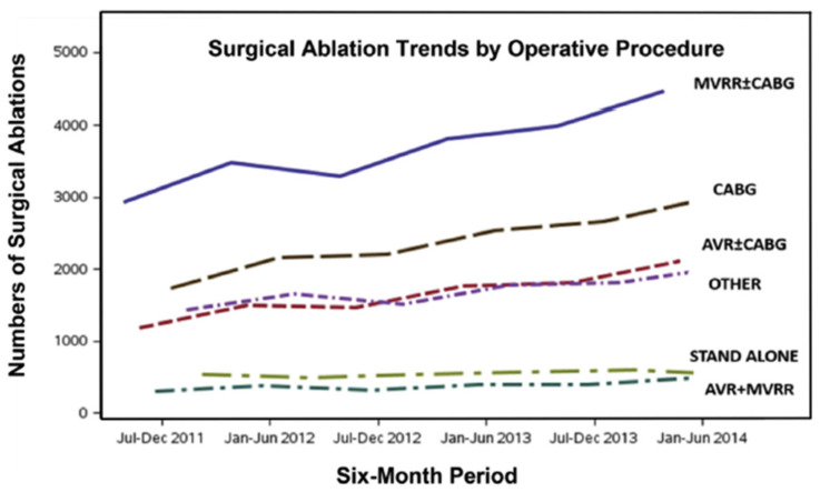
    1. Badhwar V., Rankin J.S., Ad N., Grau-Sepulveda M., Damiano R.J., Gillinov A.M., McCarthy P.M., Thourani V.H., Suri R.M., Jacobs J.P., et al. Surgical Ablation of Atrial Fibrillation in the United States: Trends and Propensity Matched Outcomes. Ann. Thorac. Surg. 2017;104:493–500. doi: 10.1016/j.athoracsur.2017.05.016. - DOI - PubMed

LinkOut - more resources