. 2022 Jan 10;23(1):4.
doi: 10.1186/s10194-021-01383-8.

Migraine-relevant sex-dependent activation of mouse meningeal afferents by TRPM3 agonists

Affiliations

Migraine-relevant sex-dependent activation of mouse meningeal afferents by TRPM3 agonists

G Krivoshein et al. J Headache Pain. .

Erratum in

Abstract

Background: Migraine is a common brain disorder that predominantly affects women. Migraine pain seems mediated by the activation of mechanosensitive channels in meningeal afferents. Given the role of transient receptor potential melastatin 3 (TRPM3) channels in mechanical activation, as well as hormonal regulation, these channels may play a role in the sex difference in migraine. Therefore, we investigated whether nociceptive firing induced by TRPM3 channel agonists in meningeal afferents was different between male and female mice. In addition, we assessed the relative contribution of mechanosensitive TRPM3 channels and that of mechanosensitive Piezo1 channels and transient receptor potential vanilloid 1 (TRPV1) channels to nociceptive firing relevant to migraine in both sexes.

Methods: Ten- to 13-week-old male and female wildtype (WT) C57BL/6 J mice were used. Nociceptive spikes were recorded directly from nerve terminals in the meninges in the hemiskull preparations.

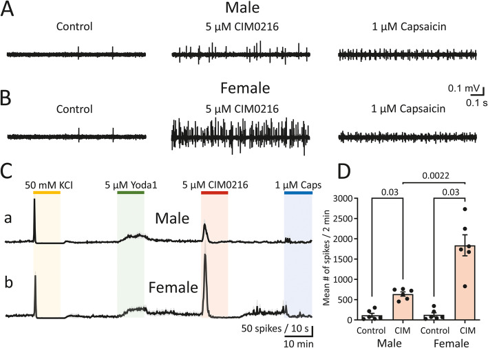
Results: Selective agonists of TRPM3 channels profoundly activated peripheral trigeminal nerve fibres in mouse meninges. A sex difference was observed for nociceptive firing induced by either PregS or CIM0216, both agonists of TRPM3 channels, with the induced firing being particularly prominent for female mice. Application of Yoda1, an agonist of Piezo1 channels, or capsaicin activating TRPV1 channels, although also leading to increased nociceptive firing of meningeal fibres, did not reveal a sex difference. Cluster analyses of spike activities indicated a massive and long-lasting activation of TRPM3 channels with preferential induction of large-amplitude spikes in female mice. Additional spectral analysis revealed ​a dominant contribution of spiking activity in the α- and β-ranges following TRPM3 agonists in female mice.

Conclusions: Together, we revealed a specific mechanosensitive profile of nociceptive firing in females and suggest TRPM3 channels as a potential novel candidate for the generation of migraine pain, with particular relevance to females.

Keywords: CIM0216; Migraine; Nociception; Pregnenolone sulfate; Sex-dependence; TRPM3.


Conflict of interest statement

The authors declare that they have no competing interests.

Figures

Fig. 1
Fig. 1
Activation of trigeminal nerve terminals by PregS in male and female mouse meninges. (A, B) Example traces of multi-unit activity (MUA) in the TG nerve innervating meninges in a hemiskull preparation of male (A) and female(B) mice recorded in the control condition (left), after application 5 μM Yoda1 (middle) and after 50 μM PregS (right). (C) Time course of spike frequency (10-s bin size) induced by application of 50 mM KCl, 5 μM Yoda1, 50 μM PregS and 1 μM capsaicin in males and females (mean ± SEM, n = 5 and n = 7, respectively). Notice the stable recovery of persistence firing during a 20-min washout after each drug application. (D) Histograms show the mean number of nociceptive spikes during 10-min recordings before (control) and after application of 50 μM PregS in males (mean ± SEM, n = 5, p = 0.06, Wilcoxon signed-rank test) and females (mean ± SEM, n = 7, p = 0.01, Wilcoxon signed-rank test). Notice the sex difference in firing when 50 μM PregS was applied (p = 0.0025, Mann–Whitney U test)
Fig. 2
Fig. 2
Activation of Piezo1 channels has no impact on increased nociceptive firing during PregS application. (A) Time course of MUA in meningeal nerves of female mice when first 5 μM Yoda1 and then 50 μM PregS was applied (mean ± SEM, n = 7). (B) Time course of MUA in female mice when first 50 μM PregS and then 5 μM Yoda1 was applied (mean ± SEM, n = 5). (C) Histograms show the percentage of increased nociceptive firing in females during 10-min recordings following 50 μM PregS application when administered before (mean ± SEM, n = 5) and after 5 μM Yoda1 application (mean ± SEM, n = 7). Notice that there is no difference in the proportion of increased nociceptive firing between the different orders of drug application (p = 0.63, Mann–Whitney U test). (D) Histograms show the percentage of increased nociceptive firing in females during 10-min recordings following 5 μM Yoda1 application when administered before (mean ± SEM, n = 7) and after 50 μM PregS application (mean ± SEM, n = 5). Notice there is no difference in the fraction of increased nociceptive firing between the different orders of drug application (p = 0.53, Mann–Whitney U test)
Fig. 3
Fig. 3
Activation of trigeminal nerve terminals by CIM0216 in male and female mouse meninges. (A, B) Example traces of MUA in TG nerves of male (A) and female (B) mice recorded in the control condition (left), after application 5 μM CIM0216 (middle) and after 1 μM capsaicin (right). (C) Time course of spike frequency induced by application of 50 mM KCl, 5 μM Yoda1, 5 μM CIM0216 and 1 μM capsaicin in males and females (mean ± SEM, 10-s bin size, n = 6 for both sexes). Notice the difference in the 2-min active phase of 5 μM CIM0216 that was much more pronounced in females, and the subsequent silent time in males compared to overactivated firing recovery during the 20-min washout period in females. (D) Histograms show the mean number of nociceptive spikes during 10-min recordings in the control condition and after application of 5 μM CIM0216 in males (mean ± SEM, n = 6, p = 0.03, Wilcoxon signed-rank test) and females (mean ± SEM, n = 6, p = 0.03, Wilcoxon signed-rank test). Notice the sex difference in firing when 5 μM CIM0216 was applied (p = 0.0022, Mann–Whitney U test)
Fig. 4
Fig. 4
No sex-difference in general nociceptive fibre excitability, Piezo1 activity and capsaicin sensitivity. (A) Histograms show the mean ± SEM number of nociceptive spikes in control conditions in males (n = 11) and females (n = 13) and the lack of a sex difference (p = 0.78, Mann–Whitney U test). (B) Histograms show the mean ± SEM number of nociceptive spikes during the 30-s active phase of 50 mM KCL application in males (n = 11) and females (n = 13) and the lack of a sex difference (p = 0.29, Mann–Whitney U test). (C) Histograms show the mean ± SEM number of nociceptive spikes during 10-min recordings after application of 5 μM Yoda1 in males (n = 11) and females (n = 13). (D) Histograms show the mean ± SEM number of nociceptive spikes during the 2-min active phase of 1 μM capsaicin application in males (n = 5) and females (n = 7). Notice the lack of a sex difference on firing when 5 μM Yoda (p = 0.94, Mann–Whitney U test) and 1 μM capsaicin (p = 0.53, Mann–Whitney U test) was applied
Fig. 5
Fig. 5
Co-activation of TRPM3 mechanosensitive receptors, Piezo1 mechanosensitive receptors, and TRPV1 channels. (A) Presentation of spike clusters within one experiment. Spikes are plotted by separating negative vs. positive amplitude in the control condition (top panels) and after 50 μM PregS application (bottom panels) in females (left) and males (right). Each individual dot indicates a single spike. Dots with similar colours represent one cluster (i.e. representing one fibre or one group of spikes, as separated by the KlustaKwik method. (B, C) Examples of clusters within one nerve of female (B) and male (C) meninges that were sequentially activated by all three agonists (Ba, Ca), both 5 μM Yoda1 and 50 μM PregS (Bb, Cb) or both 50 μM PregS and 1 μM capsaicin (Bc, Cc), or only 50 μM PregS (Bd, Cd). Spike shapes are depicted for each cluster. (D, E) Pie diagrams demonstrate the averaged percentage of clusters with various neurochemical profiles in females (D) and males (E). Data are presented from 7 and 5 experiments in females and males, respectively. Notice, the difference in co-appearance of responses to 5 μM Yoda1 and 50 μM PregS in females and males. In females, up to 44% clusters were activated by PregS and Yoda1 (23%) or by PregS, Yoda1 and capsaicin (21%), whereas in males, the number of such ‘supermechanosensitive’ fibres was only 24% (PregS and Yoda1 (7%); PregS, Yoda1 and capsaicin (17%)). In males, 7% of clusters showed no response to any agonist, whereas this was the case for only 1% of clusters in females where, in addition, no fibres responded only to 5 μM Yoda1
Fig. 6
Fig. 6
Spectral analysis of nociceptive firing in control conditions and the presence of CIM0216. (A, B) Example traces of MUA within one experiment in the control condition and during 5 μM CIM0216 in females (A) and males (B). Notice, there is only a single spike in the control condition in both males and females, while during the 2-min active phase of 5 μM CIM0216 application, the number of spikes increased differently for females and males, within the initial 0.5-s of drug application. (C, D) Spectrograms show the average interspike intervals (ISI) for all experiments in females (C) in the control conditions (grey line, n = 13), after application of 50 μM PregS (blue line, n = 7) and after 5 μM CIM0216 application (red line, n = 6) and in males (D) in the control condition (grey line, n = 11), after application of 50 μM PregS (blue line, n = 5) and after 5 μM CIM0216 application (red line, n = 6) on panel. The dotted vertical line at 0.1 ISI indicates the 10 Hz frequency. The number of ISIs below 0.1 s (i.e. reflecting spiking activity above 10 Hz frequency) is higher in females after 50 μM PregS and 5 μM CIM0216. (E, F) Distribution of fibres (clusters) according to the firing frequency in the control condition (top panel; grey) and after application of 5 μM CIM0216 (bottom panel; red) presented as mean ± SEM for females (E) (n = 6) and males (F) (n = 6) on panel (F). Notice that 5 μM CIM0216 induced nociceptive firing with θ-range of spiking activity for both sexes, whereas in females, θ-range, as well as higher spiking activity of α and β-ranges, were induced

References

    1. Global Burden of Disease Study (2019) US Seattle, Institute for Health Metrics and Evaluation http://ghdxhealthdataorg/gbd-results-tool Accessed 15 Oct 2020
    1. Launer LJ, Terwindt GM, Ferrari MD. The prevalence and characteristics of migraine in a population-based cohort. Neurology. 1999;53(3):3–537. doi: 10.1212/WNL.53.3.537. - DOI - PubMed
    1. Headache Classification Committee of the International Headache Society (IHS) The international classification of headache disorders, 3rd edition. Cephalalgia. 2018;38(1):1–211. doi: 10.1177/0333102417738202. - DOI - PubMed
    1. MacGregor EA. Menstrual and perimenopausal migraine: a narrative review. Maturitas. 2020;142:24–30. doi: 10.1016/j.maturitas.2020.07.005. - DOI - PubMed
    1. Finocchi C, Ferrari M. Female reproductive steroids and neuronal excitability. Neurol Sci. 2011;32(S1):31–35. doi: 10.1007/s10072-011-0532-5. - DOI - PubMed