. 2022 Jan 10;20(1):29.
doi: 10.1186/s12951-021-01206-7.

Bone mesenchymal stem cell-derived exosomal microRNA-7-5p inhibits progression of acute myeloid leukemia by targeting OSBPL11

Affiliations

Bone mesenchymal stem cell-derived exosomal microRNA-7-5p inhibits progression of acute myeloid leukemia by targeting OSBPL11

Duanfeng Jiang et al. J Nanobiotechnology. .

Retraction in

Abstract

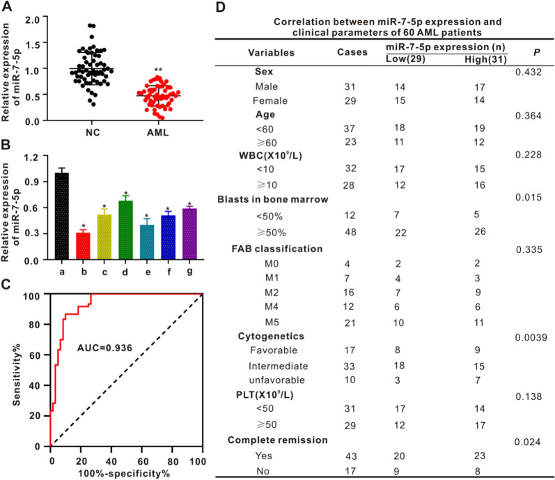
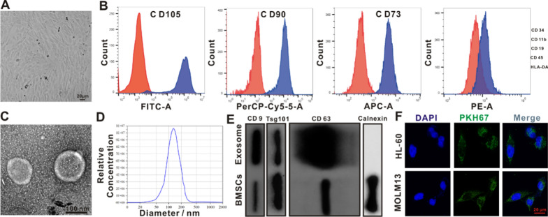
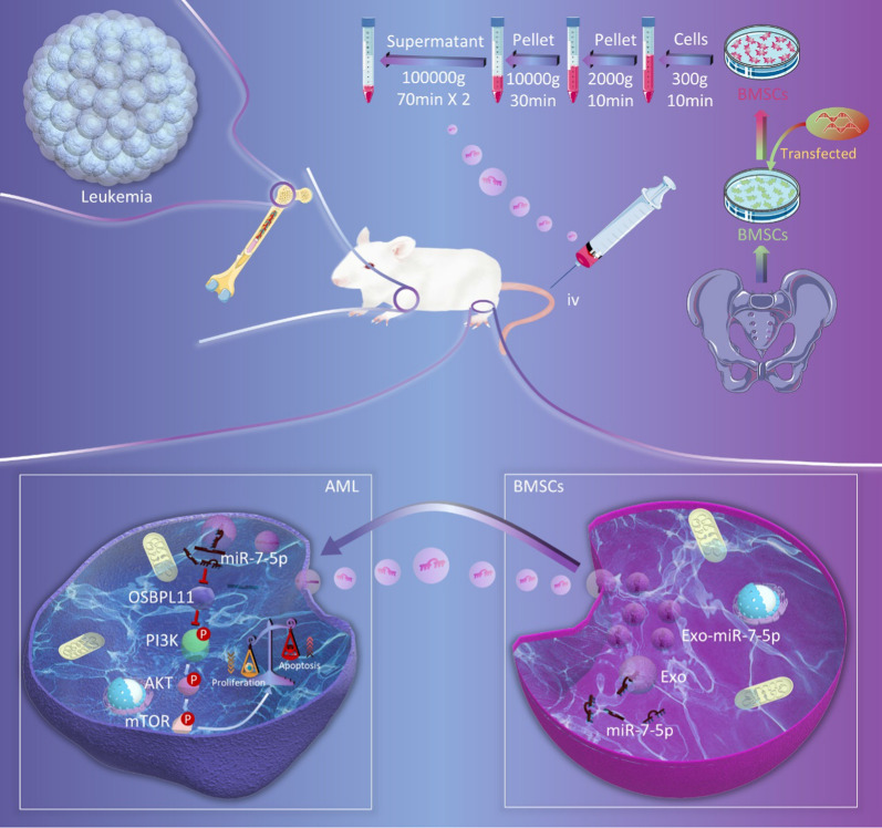
Background: Acute myeloid leukemia (AML) is a malignant clonal disease of hematopoietic stem- and progenitor-cell origin. AML features massive proliferation of abnormal blasts and leukemia cells in the bone marrow and the inhibition of normal hematopoiesis at onset. Exosomes containing proteins or nucleic acids are secreted by cells; they participate in intercellular communication and serve as key modulators of hematopoiesis. The purpose of this study was to investigate the effects of exosomes derived from bone marrow mesenchymal stem cells (BMSCs) on the regulation of AML and the underlying mechanisms mediated by microRNA (miRNA).

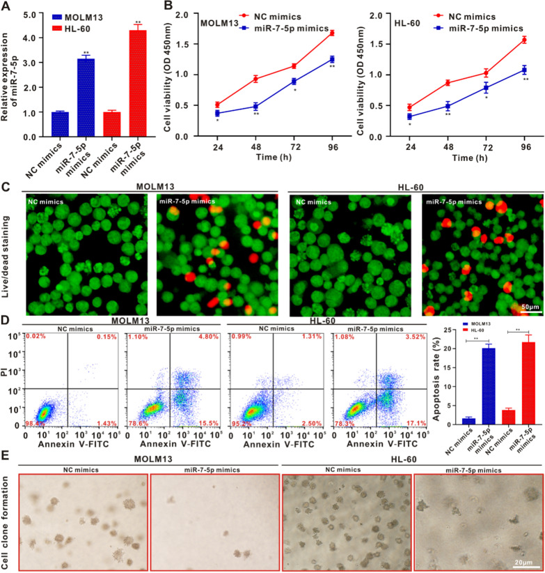
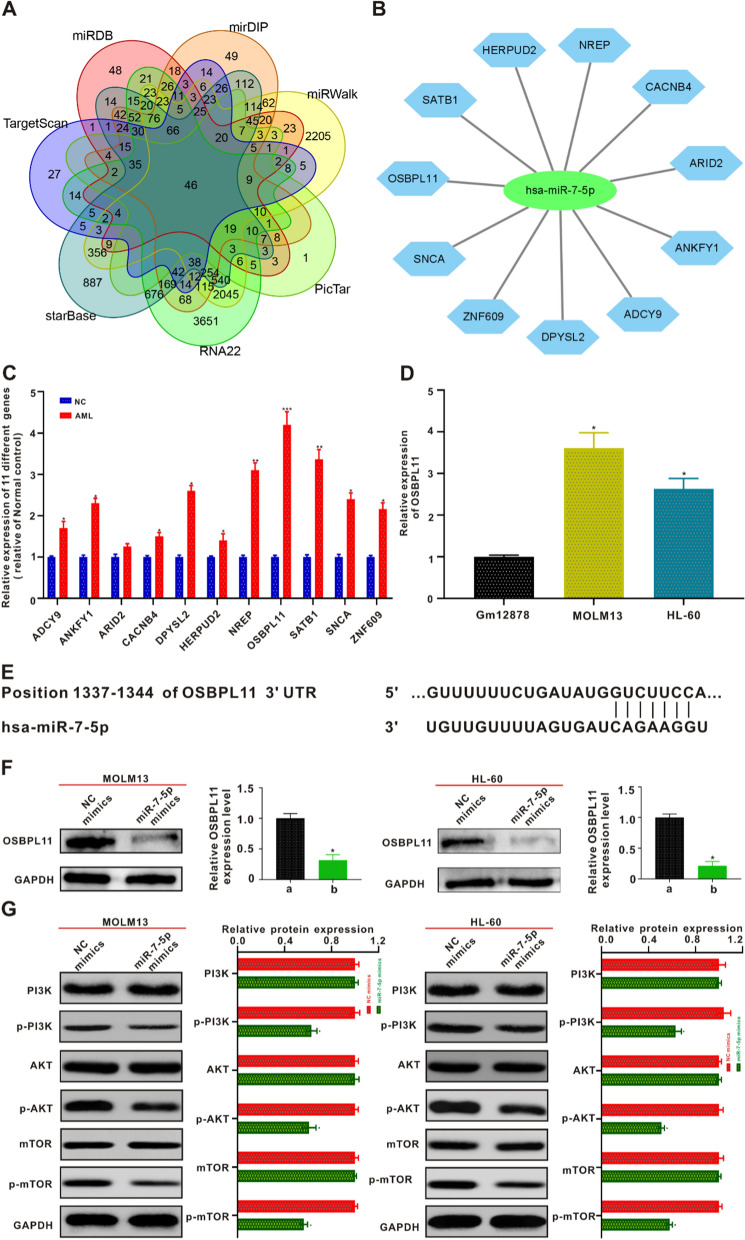
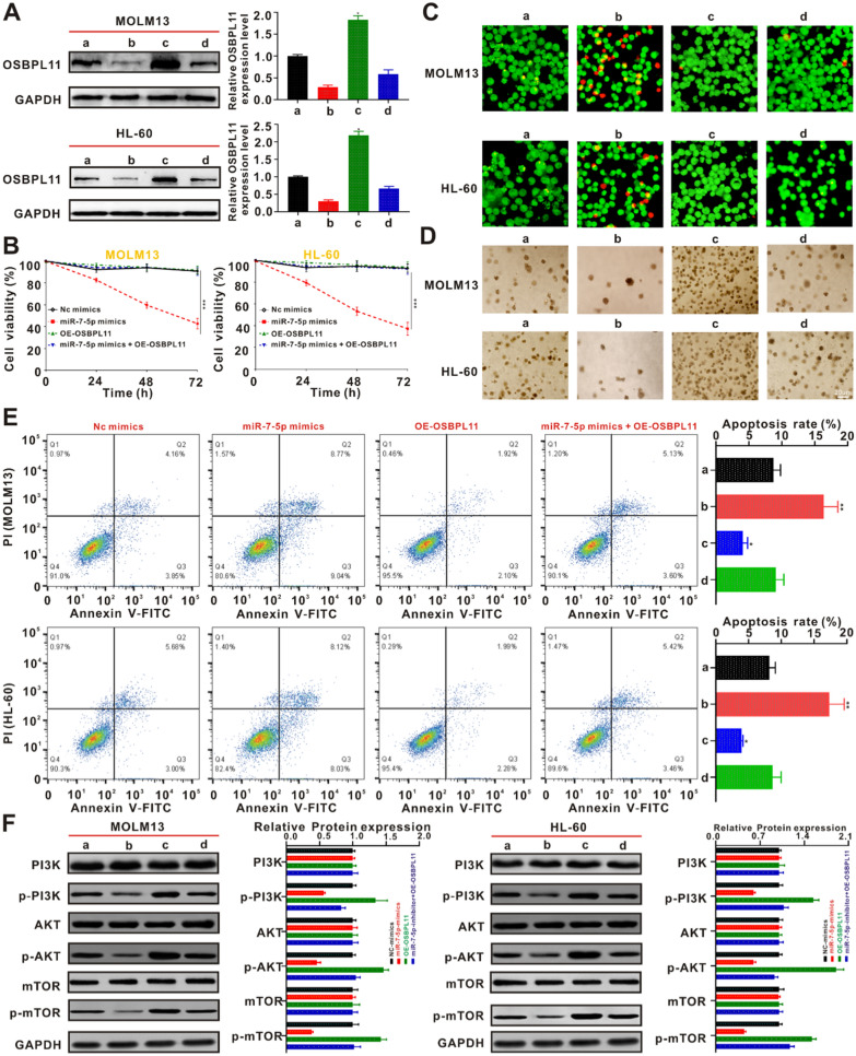
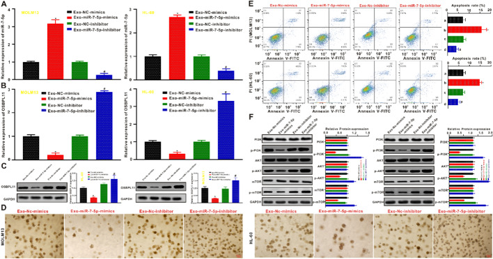
Methods: Dysregulated miR-7-5p in AML patients was identified using qRT-PCR and its clinical significance was explored. Bioinformatic analysis revealed the target gene OSBPL11 that could be regulated by miR-7-5p. The findings were validated using a dual-luciferase reporter assay and western blotting. The functional genes of the PI3K/AKT/mTOR signaling pathway were identified, and the functional significance of miR-7-5p in AML cells was determined using a functional recovery assay. AML cells were co-cultured with exosomes originating from BMSCs overexpressing miR-7-5p to determine cell-cell regulation by Exo-miR-7-5p, as well as in vitro and in vivo functional validation via gain- and loss-of-function methods.

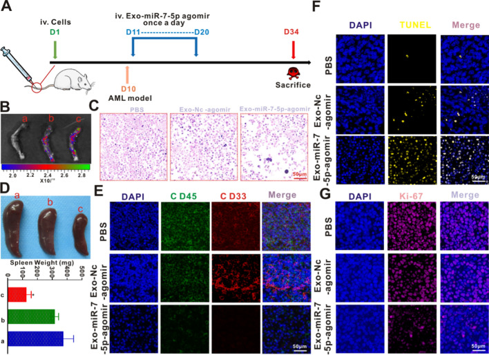
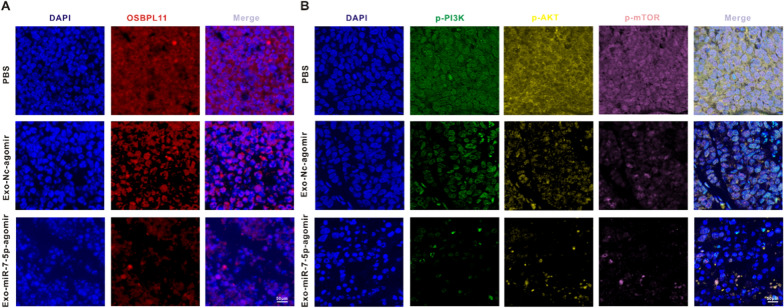
Results: Expression of miR-7-5p was decreased in AML patients and cells. Overexpression of miR-7-5p curbed cellular proliferation and promoted apoptosis. Overexpression of OSBPL11 reversed the tumorigenic properties of miR-7-5p in AML cells in vitro. Exo-miR-7-5p derived from BMSCs induced formation of AML cells prone to apoptosis and a low survival rate, with OSBPL11 expression inhibited through the PI3K/AKT/mTOR signaling pathway. Exo-miR-7-5p derived from BMSCs exhibited tumor homing effects in vitro and in vivo, and inhibited AML development.

Conclusions: Exo-miR-7-5p derived from BMSCs negatively regulates OSBPL11 by suppressing the phosphorylation of the PI3K/AKT/mTOR signaling pathway, thereby inhibiting AML proliferation and promoting apoptosis. The data will inform the development of AML therapies based on BMSC-derived exosomes.

Keywords: Acute myeloid leukemia; Bone marrow mesenchymal stem cells; Exosome; OSBPL11; PI3K/AKT/mTOR; miR-7-5p.


Conflict of interest statement

The authors declare that they have no competing interests.

Figures

Fig. 1
Fig. 1
The expression of MiR-7-5p is low in the peripheral blood of AML patients and AML cell lines. A The expressing of miR-7-5p was remarkably reduced in the peripheral blood of AML sufferers (n = 60) compared with that of the Normal Controls (NC) (n = 60). **p < 0.01 in contrast to NC. B MiR-7-5p expressing in human normal hematopoietic cell GM12878 (a) and the AML cell lines MOLM13 (b), THP1 (c), U937 (d), HL60 (e), MV4-11 (f) and KG-1 (g). *p < 0.05 compared with GM12878. C ROC curve analysis revealed that miR-7-5p was an underlying marker for screening AML patients from healthy controls. D Correlation of the peripheral blood miR-7-5p level with clinical characteristics of 60 AML patients
Fig. 2
Fig. 2
MiR-7-5p suppresses the proliferative activity and promotes the apoptosis of MOLM13 and HL-60 cells. A qRT-PCR analysis verifying the transfection efficiency of the miR-7-5p mimics. B MOLM13 and HL-60 cells were cultivated in the intermediary with/without miR-7-5p mimics for 24, 48, 72 and 96 h. The survival rate of the cells was identified by CCK-8 analysis. *p < 0.05 in contrast to the NC mimic group, **p < 0.01 in contrast to the NC mimic group. C Live/dead staining of MOLM13 and HL-60 cells upon with/without miR-7-5p mimics for 24 h. Scale bar: 50 μm. D Flow cytometry was used to identify the programmed cell death of MOLM13 and HL-60 cells posterior to 24 h. Data are described as the average ± SD (n = 3). (**p < 0.01). (E) Typical pictures of the colony formation of MOLM13 and HL-60 cells treated with/without miR-7-5p mimics for 14 days
Fig. 3
Fig. 3
MiR-7-5p targeted gene prediction and the validation. A Seven network data bases forecast the number of targeted genes of miR-7-5p and take their intersection using Venn diagram. (B) A network map between 11 target genes and miR-7-5p was predicted. C qRT-PCR was employed to validate the expressing levels of 11 predicted targeted genes in AML patients and NC. *p < 0.05 in contrast to the NC, **p < 0.01 in contrast to the NC, ***p < 0.001 in contrast to the NC. D Expression of OSBPL11 mRNA in AML cells and human hematopoietic normal cells GM12878 by qRT-PCR. E The sequencing result of mankind miR-7-5p and the forecasted binding areas with miR-7-5p in the OSBPL11 non-translated area (3′-UTR) are displayed. F After co-culturing with MOLM13 and HL-60 cells with miR-7-5p mimics for 24 h, respectively, OSBPL11 protein expression levels were measured by immunoblotting. The quantitation data from immunoblotting analysis were analyzed via ImageJ program. G MiR-7-5p mimics were co-cultured with MOLM13 and HL-60 cells for 24 h, and PI3K, p-PI3K, AKT, p-AKT, mTOR and p-mTOR protein expressing status were identified by western blotting, respectively. The quantitation data from immunoblotting analysis were analyzed via ImageJ program, which were described as mean ± SD (n = 3)
Fig. 4
Fig. 4
Overexpression of OSBPL11 blocked the miR-7-5p-mediated potency on the apoptosis in AML cells. A MOLM13 and HL-60 cells were subjected to NC mimics transfection (a), miR-7-5p mimics (b), OE-OSBPL11 (c) and miR-7-5p mimics + OE-OSBPL11 (d), respectively, and then co-cultured for 24 h. OSBPL11 protein expression levels were measured by immunoblotting. The quantitation data from immunoblotting analysis were analyzed via ImageJ program. B OSBPL11 expression partly counteract the miR-7-5p-mediated suppressive potency on the proliferative activity of MOLM13 and HL-60 cells. C Live/dead staining of MOLM13 and HL-60 cells under different treating for 24 h. Plotting scale: 50 μm. a NC mimics, b miR-7-5p mimics, c OE-OSBPL11, d miR-7-5p mimics + OE-OSBPL11. D Representative images of colony formation of MOLM13 and HL-60 cells treated with a NC mimics, b miR-7-5p mimics, c OE-OSBPL11, d miR-7-5p mimics + OE-OSBPL11 for 14 days, respectively. E Flow cytometry was employed to identify the programmed cell death of MOLM13 and HL-60 cells after 24 h. Data are expressed as the average ± SD (n = 3). *p < 0.05 in contrast to the NC mimics, **p < 0.01 in contrast to the NC mimics. F a NC mimics, b miR-7-5p mimics, c OE-OSBPL11, d miR-7-5p mimics + OE-OSBPL11 were co-cultured with MOLM13 and HL-60 cells for 24 h, protein expression of PI3K/AKT/mTOR signal path biomarkers were identified via immunoblotting. The quantitation data from immunoblotting analysis were analyzed via ImageJ program, which were expressed as mean ± SD (n = 3)
Fig. 5
Fig. 5
Isolation and characterization of exosomes from BMSCs. A Representative morphology of BMSCs. B Flow cytometry assay revealed that BMSCs were positive for mesenchyma lineage biomarkers (CD73, CD90 and CD105), negative for hematopoiesis and endothelium biomarkers (CD34, CD11b, CD19, CD45), and negative for HLA-DR. C TEM presented the morphological status of BMSCs-originated exosomes. Scale bar:100 nm. D Nanoparticle tracing assay of the BMSCs-originated exosomes. E Immunoblotting assay of superficial biomarkers CD9, CD63 and Tsg101 expressing standardized to Calnexin in the BMSCs-originated exosomes. F Images of BMSCs-exosomes labeled with PKH-67 (green) taken up by MOLM13 and HL-60 were analyzed via CLSM. Scale bar: 20 μm
Fig. 6
Fig. 6
BMSCs-derived exosome miR-7-5p inhibits tumorigenicity of AML in vitro. A The identification of miR-7-5p expressing posterior to the transfection by qRT-PCR. B The identification of OSBPL11 mRNA expressing after transfection by qRT-PCR. C Exo-miR-7-5p mimics and Exo-miR-7-5p inhibitor were co-cultured with MOLM13 and HL-60 cells, respectively, and OSBPL11 protein expression after transfection was determined by Western blotting. D Representative images of colony formation of MOLM13 and HL-60 cells subjected to Exo-miR-7-5p mimics treatment or Exo-miR-7-5p inhibitor for 14 days, respectively. E Flow cytometry was employed to identify the programmed cell death of MOLM13 and HL-60 cells posterior to 24 h. Data are expressed as the average ± SD (n = 3). F Exo-miR-7-5p mimics or Exo-miR-7-5p inhibitor were co-cultured with MOLM13 and HL-60 cells for 24 h, protein expression of PI3K/AKT/mTOR signal path biomarkers were identified via immunoblotting. The quantitation data from immunoblotting analysis were analyzed via ImageJ program, which were expressed as average ± SD (n = 3). *p < 0.05 in contrast to the Exo-Nc-mimics, #p < 0.05 in contrast to the Exo-Nc-inhibitor
Fig. 7
Fig. 7
Anti-AML potency of Exo-miR-7-5p agomir in vivo. A Schematic diagram of in vivo experimental design. B Ex vivo imaging also revealed that these exosomes derived from BMSCs have active targeting properties to the bone marrow compared to PBS. a PBS, b Exo-NC agomir and c Exo-miR-7-5p agomir. C Representative images of leukemic bone marrow tissue after H&E staining of mice executed on day 34 by injecting PBS through tail vein, Exo-NC agomir and Exo-miR-7-5p agomir, respectively. Scale bar: 50 μm. D Spleen size and weight after the sacrifice of all mice on day 34 after treatment of the tested mice with a PBS, b Exo-NC agomir and c Exo-miR-7-5p agomir, respectively. E Tumor burden (CD45 and CD33) of splenic leukemic cell infiltration by immunofluorescence. F, G The animals were under the treatment of PBS, Exo-NC agomir and Exo-miR-7-5p agomir, respectively, and TUNEL (yellow) and Ki-67 (pink) of the bone marrow group were detected by immunofluorescence after the mice were executed on day 34. Scale bar: 50 μm
Fig. 8
Fig. 8
Mechanistic validation of BMSCs-derived exosome miR-7-5p-agomir against AML in vivo. A OSBPL11 (red) in the leukemic bone marrow tissues was analyzed by immunofluorescence staining after the mice were treated with different treatment modalities. Scale bar: 50 μm. B The animals were under the treatment of PBS, Exo-NC agomir and Exo-miR-7-5p agomir, respectively, and p-PI3K (green), p-AKT (yellow) and p-mTOR (pink) in the leukemic bone marrow tissues were analyzed by immunofluorescence staining. Scale bar: 50 μm
Fig. 9
Fig. 9
Schematic diagram of miR-7-5p mimics transfected into BMSCs. The extracted exosome miR-7-5p inhibits leukemia proliferation and promotes apoptosis by negatively regulating OSBPL11 and inhibiting PI3K/AKT/mTOR signaling pathway phosphorylation

References

    1. Khaldoyanidi S, Nagorsen D, Stein A, Ossenkoppele G, Subklewe M. Immune biology of acute myeloid leukemia: implications for immunotherapy. J Clin Oncol. 2021;39:419–32. - DOI - PMC - PubMed
    1. Du A, Wu X, Gao Y, Jiang B, Wang J, Zhang P, Zhao Q. m6A regulator-mediated methylation modification patterns and tumor microenvironment infiltration characterization in acute myeloid leukemia. Front Immunol. 2021;12:789914. - DOI - PMC - PubMed
    1. Short NJ, Konopleva M, Kadia TM, Borthakur G, Ravandi F, DiNardo CD, Daver N. Advances in the treatment of acute myeloid leukemia: new drugs and new challenges. Cancer Discov. 2020;10:506–25. - DOI - PubMed
    1. Yang X, Wang J. Precision therapy for acute myeloid leukemia. J Hematol Oncol. 2018;11:3. - DOI - PMC - PubMed
    1. Duncavage EJ, Schroeder MC, O’Laughlin M, Wilson R, MacMillan S, Bohannon A, Kruchowski S, Garza J, Du F, Hughes AEO, et al. Genome sequencing as an alternative to cytogenetic analysis in myeloid cancers. N Engl J Med. 2021;384:924–35. - DOI - PMC - PubMed

Publication types