. 2022 Jan 10;15(1):7.
doi: 10.1186/s13104-021-05896-y.

Carotid artery velocity time integral and corrected flow time measured by a wearable Doppler ultrasound detect stroke volume rise from simulated hemorrhage to transfusion

Affiliations

Carotid artery velocity time integral and corrected flow time measured by a wearable Doppler ultrasound detect stroke volume rise from simulated hemorrhage to transfusion

Jon-Émile S Kenny et al. BMC Res Notes. .

Abstract

Objective: Doppler ultrasonography of the common carotid artery is used to infer stroke volume change and a wearable Doppler ultrasound has been designed to improve this workflow. Previously, in a human model of hemorrhage and resuscitation comprising approximately 50,000 cardiac cycles, we found a strong, linear correlation between changing stroke volume, and measures from the carotid Doppler signal, however, optimal Doppler thresholds for detecting a 10% stroke volume change were not reported. In this Research Note, we present these thresholds, their sensitivities, specificities and areas under their receiver operator curves (AUROC).

Results: Augmentation of carotid artery maximum velocity time integral and corrected flowtime by 18% and 4%, respectively, accurately captured 10% stroke volume rise. The sensitivity and specificity for these thresholds were identical at 89% and 100%. These data are similar to previous investigations in healthy volunteers monitored by the wearable ultrasound.

Keywords: Carotid Doppler; Corrected flow time; Stroke volume; Velocity time integral.


Conflict of interest statement

JESK, ME, ZY, AME, JKE are employees of Flosonics Medical, a start-up building the wearable ultrasound, IB reports consulting fees for GE Healthcare, DCM, CHK and BDJ report no conflicts.

Figures

Fig. 1
Fig. 1
Picture of wireless, wearable Doppler ultrasound device
Fig. 2
Fig. 2
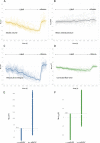
Hemodynamic data captured during lower body negative pressure (LBNP) and release. Measures from AD are synchronously captured. Each faint line represents a single protocol, while the emboldened line represents the average of all protocols. A Stroke volume (SV) percent change during progressively severe LBNP (i.e., hemorrhage model) and release of LBNP (i.e., rapid transfusion model). B Mean arterial pressure (MAP) percent change. C velocity time integral (VTI) from the wearable Doppler percent change. D corrected flow time (ccFT) percent change. E The optimal carotid artery maximum VTI threshold for distinguishing ≥  + 10% SV. Each data point represents a 10-s average. Prior to subsampling, there were 3596 data points categorized as <  + 10% SV and 598 data points categorized as ≥  + 10% SV. The data categorized as <  + 10% SV were randomly subsampled, iteratively 1000 times, to 598 data points (see methods). The sensitivity of maximum VTI is 532/598 = 89% and specificity is 598/598 = 100%. F The optimal ccFT threshold for distinguishing + 10% SV. Each data point represents a 10-s average. The sensitivity is 532/598 = 89% and specificity is 598/598 = 100%

References

    1. Kenny J-ES, Barjaktarevic I. Letter to the editor: stroke volume is the key measure of fluid responsiveness. Crit Care. 2021;25(1):104. doi: 10.1186/s13054-021-03498-5. - DOI - PMC - PubMed
    1. Kenny J-ÉS. Functional hemodynamic monitoring with a wireless ultrasound patch. J Cardiothorac Vasc Anesth. 2021;35(5):1509–1515. doi: 10.1053/j.jvca.2021.01.040. - DOI - PubMed
    1. Marik PE, Levitov A, Young A, Andrews L. The use of bioreactance and carotid Doppler to determine volume responsiveness and blood flow redistribution following passive leg raising in hemodynamically unstable patients. Chest. 2013;143(2):364–370. doi: 10.1378/chest.12-1274. - DOI - PubMed
    1. Barjaktarevic I, Toppen WE, Hu S, Montoya EA, Ong S, Buhr R, et al. Ultrasound assessment of the change in carotid corrected flow time in fluid responsiveness in undifferentiated shock. Crit Care Med. 2018;11:1040–1046. doi: 10.1097/CCM.0000000000003356. - DOI - PMC - PubMed
    1. Beier L, Davis J, Esener D, Grant C, Fields JM. Carotid ultrasound to predict fluid responsiveness: a systematic review. J Ultrasound Med. 2020;39(10):1965–1976. doi: 10.1002/jum.15301. - DOI - PubMed