

Applications of the amniotic membrane in tissue engineering and regeneration: the hundred-year challenge
- PMID: 35012669
- PMCID: PMC8744057
- DOI: 10.1186/s13287-021-02684-0
Applications of the amniotic membrane in tissue engineering and regeneration: the hundred-year challenge
Abstract
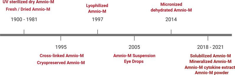
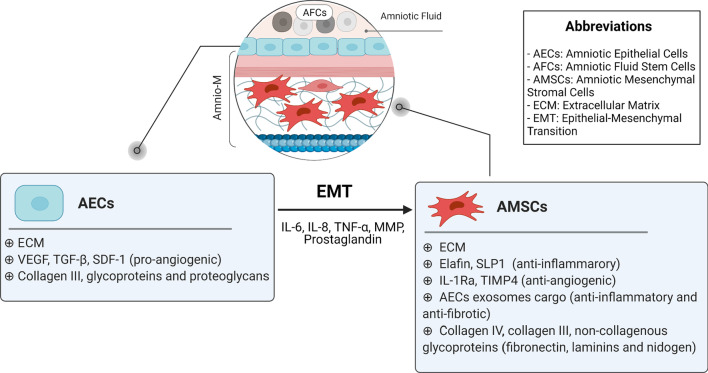
The amniotic membrane (Amnio-M) has various applications in regenerative medicine. It acts as a highly biocompatible natural scaffold and as a source of several types of stem cells and potent growth factors. It also serves as an effective nano-reservoir for drug delivery, thanks to its high entrapment properties. Over the past century, the use of the Amnio-M in the clinic has evolved from a simple sheet for topical applications for skin and corneal repair into more advanced forms, such as micronized dehydrated membrane, amniotic cytokine extract, and solubilized powder injections to regenerate muscles, cartilage, and tendons. This review highlights the development of the Amnio-M over the years and the implication of new and emerging nanotechnology to support expanding its use for tissue engineering and clinical applications.
Keywords: Amnion; Biodegradability; Natural biomaterial; Regenerative medicine; Tissue engineering.
© 2021. The Author(s).
Conflict of interest statement
No potential conflicts of interest were disclosed.
Figures


References
-
- Manuelpillai U, Moodley Y, Borlongan CV, Parolini O. Amniotic membrane and amniotic cells: potential therapeutic tools to combat tissue inflammation and fibrosis? Placenta. 2011;32(Suppl 4):S320–S325. - PubMed
-
- Salah RA, Elkhenany H, El-Badri N. Scaffold engineering using the amniotic membrane. In: El-Badri N, editor. Regenerative medicine and stem cell biology. Cham: Springer; 2020. pp. 323–346.
-
- Díaz-Prado S, Muiños-López E, Hermida-Gómez T, Cicione C, Rendal-Vázquez ME, Fuentes-Boquete I, et al. Human amniotic membrane as an alternative source of stem cells for regenerative medicine. Differentiation. 2011;81(3):162–171. - PubMed
-
- Farhadihosseinabadi B, Farahani M, Tayebi T, Jafari A, Biniazan F, Modaresifar K, et al. Amniotic membrane and its epithelial and mesenchymal stem cells as an appropriate source for skin tissue engineering and regenerative medicine. Artif Cells Nanomed Biotechnol. 2018;46(sup2):431–440. - PubMed
Publication types
MeSH terms
LinkOut - more resources
Full Text Sources
Other Literature Sources
Miscellaneous
