. 2022 Mar 1;98(9):e912-e923.
doi: 10.1212/WNL.0000000000013278. Epub 2022 Jan 10.

Diagnosis of Genetic White Matter Disorders by Singleton Whole-Exome and Genome Sequencing Using Interactome-Driven Prioritization

Collaborators, Affiliations

Diagnosis of Genetic White Matter Disorders by Singleton Whole-Exome and Genome Sequencing Using Interactome-Driven Prioritization

Agatha Schlüter et al. Neurology. .

Abstract

Background and objectives: Genetic white matter disorders (GWMD) are of heterogeneous origin, with >100 causal genes identified to date. Classic targeted approaches achieve a molecular diagnosis in only half of all patients. We aimed to determine the clinical utility of singleton whole-exome sequencing and whole-genome sequencing (sWES-WGS) interpreted with a phenotype- and interactome-driven prioritization algorithm to diagnose GWMD while identifying novel phenotypes and candidate genes.

Methods: A case series of patients of all ages with undiagnosed GWMD despite extensive standard-of-care paraclinical studies were recruited between April 2017 and December 2019 in a collaborative study at the Bellvitge Biomedical Research Institute (IDIBELL) and neurology units of tertiary Spanish hospitals. We ran sWES and WGS and applied our interactome-prioritization algorithm based on the network expansion of a seed group of GWMD-related genes derived from the Human Phenotype Ontology terms of each patient.

Results: We evaluated 126 patients (101 children and 25 adults) with ages ranging from 1 month to 74 years. We obtained a first molecular diagnosis by singleton WES in 59% of cases, which increased to 68% after annual reanalysis, and reached 72% after WGS was performed in 16 of the remaining negative cases. We identified variants in 57 different genes among 91 diagnosed cases, with the most frequent being RNASEH2B, EIF2B5, POLR3A, and PLP1, and a dual diagnosis underlying complex phenotypes in 6 families, underscoring the importance of genomic analysis to solve these cases. We discovered 9 candidate genes causing novel diseases and propose additional putative novel candidate genes for yet-to-be discovered GWMD.

Discussion: Our strategy enables a high diagnostic yield and is a good alternative to trio WES/WGS for GWMD. It shortens the time to diagnosis compared to the classical targeted approach, thus optimizing appropriate management. Furthermore, the interactome-driven prioritization pipeline enables the discovery of novel disease-causing genes and phenotypes, and predicts novel putative candidate genes, shedding light on etiopathogenic mechanisms that are pivotal for myelin generation and maintenance.


Figures

Figure 1
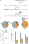
Figure 1. Diagnostic Process Diagram and Diagnostic Yield
(A) Number of cases included in the study and diagnostic process. (B) Global, whole-exome sequencing (WES), and whole-genome sequencing (WGS) diagnostic yield. (C) Percentage of diagnosis in the first WES analysis, obtained by WES reanalysis and by WGS. (D) Diagnostic percentage according to age.
Figure 2
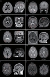
Figure 2. MRI Findings in Patients With New/Atypical and Blended Phenotypes
(A) LNF-48, 5 years. PARS2; p.Arg186Gly/p.Lys187Arg (COMP HTZ). Periatrial white matter (WM) hyperintensity (red arrows) with frontal-parietal atrophy, ventriculomegaly, and thin corpus callosum (arrowheads) (axial T2 fluid-attenuated inversion recovery [FLAIR], sagittal T1-weighted images). (B) LNF-29, 10 months. PNPT1; p.Ala507Ser (HMZ). Bilateral periatrial and temporal anterior subcortical WM hyperintensities (red arrows) with temporal cystic lesions (arrowheads) (axial T2 and coronal T2 FLAIR-weighted images). (C) LNF-47, 2 years. POLR3A; c.1771-7C > G/p.Leu1129 (COMP HTZ). Optic radiation mild WM hyperintensity (red arrows), striatal atrophy and hyperintensity (arrowheads), and superior cerebellar peduncles hyperintense signal (asterisks) (axial T2 images). (D) LNF-85, 48 years. PSEN1; p.Thr350Ile (HTZ). MRI showed diffuse WM hyperintensities (red arrows) with corpus callosum and cortical atrophy (arrowheads) (axial T2 and sagittal T1 FLAIR images). (E) LNF-88, 13 years. GFPT1; p.Asp296Val (HMZ). Axial T2 hyperintensities involving deep cerebral WM (red arrows), cerebellar peduncles (white arrows), and middle blade of corpus callosum (arrowheads), sparing subcortical WM (axial T2 and sagittal T1-weighted images). (F) LNF-114, 5 months. SCN8A; p.Val409Met (HTZ). Important myelination delay, thin corpus callosum and signs of cerebral and cerebellar atrophy (axial and sagittal T1-weighted images). (G) SPG-25, 44 years. SOX10; p.Tyr83Asp (HTZ). Periventricular WM signal abnormality, sparing U fibers (red arrows), and thin isthmus of the corpus callosum (arrowhead) (axial T2-FLAIR and sagittal T1 weighted images). (H) LNF-40.0, 13 years. CYP2U1; p.Arg178Thr (HMZ) and LNF-40.4, 15 years. PAH; p.Thr380Met (HMZ). Periventricular WM hyperintensities (red arrows) (axial T2 weighted images). (I) LNF-56, 15 years. POLR3A; p.Cys724Tyr/p.Pro705Ala (COMP HTZ) and CACNA1A; p.Tyr546Ter (HTZ). Periventricular symmetric heterogeneous WM hyperintensities (red arrows) and hypointensity in globus pallidus (arrowheads), thalamic anterolateral nuclei (asterisks), optic radiations, and pyramidal tracts, with mild atrophy of the cerebellar superior vermis (white arrow) (axial T2 and sagittal T1-weighted images). (J) LNF-89.3, 15 years. CP; p.Gly868GlufsTer26 (HMZ)/NDUFS1; p.Ser701Asn (HTZ). Periventricular symmetric T2 hyperintensity with cystic degeneration and pyramidal tract involvement (red arrows) and corpus callosum atrophy. Accumulation of paramagnetic material in the substantia nigra (asterisks) (axial T2-FLAIR and axial susceptibility-weighted imaging).
Figure 3
Figure 3. MRI of Selected Cases With Variants in Hereditary Spastic Paraparesis Genes and White Matter Involvement
T2 hyperintensity in the bilateral periventricular white matter. (A, D) Axial T2 images. (B, C) Axial T2 fluid-attenuated inversion recovery images.
Figure 4
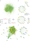
Figure 4. GWMD Expanded Interactome
(A) The genetic white matter disorder (GWMD) seeds + expanded network was generated by the network prioritization tool, resulting in 1,530 proteins. The seed genes known to be mutated in GWMD are shown in yellow circles, disease genes not previously associated with GWMD are shown in green, and new GWMD candidates are shown in blue. Comparison of statistical connectivity strength of the GWMD expanded network with 1,000 permutations of randomly selected proteins from the global human network. Red dots denote the value of the metric on the GWMD expanded network constituted by 1,530 proteins. Box and whisker plots denote matched null distributions (i.e., 1,000 permutations). (D, left) Within-group edge count (i.e., number of edges between members of the query set). (D, right) distance is the average path length in the network obtained by calculating the shortest paths between all pairs of proteins. (B–E) Zoom in the network for specific putative candidates as illustrative example of the GWMD expanded network potentiality. (B) Delta 4-desaturase, sphingolipid 1 (DEGS1); (C) phosphatidylinositol 4-kinase alpha (PI4KA); (D) mitochondrial ribosome-associated GTPase 1 (MTG1); and (E) potassium voltage-gated channel subfamily A regulatory beta subunit 2 (KCNAB2) protein. *Recently associated with leukodystrophy. White matter expanded network available in NDEx repository at public.ndexbio.org/#/network/fd5fc166-9ecc-11eb-9e72-0ac135e8bacf?accesskey=a75ac048b59aca2c9310c04a6f1d96ea34052231d9204f284c5e1d420fc2ca26

References

    1. Fogel BL, Lee H, Deignan JL, et al. . Exome sequencing in the clinical diagnosis of sporadic or familial cerebellar ataxia. JAMA Neurol. 2014;71(10):1237-1246. - PMC - PubMed
    1. Gonzaga-Jauregui C, Harel T, Gambin T, et al. . Exome sequence analysis suggests that genetic burden contributes to phenotypic variability and complex neuropathy. Cell Rep. 2015;12(7):1169-1183. - PMC - PubMed
    1. van de Warrenburg BP, Schouten MI, de Bot ST, et al. . Clinical exome sequencing for cerebellar ataxia and spastic paraplegia uncovers novel gene-disease associations and unanticipated rare disorders. Eur J Hum Genet. 2016;24(10):1460-1466. - PMC - PubMed
    1. Vanderver A, Simons C, Helman G, et al. . Whole exome sequencing in patients with white matter abnormalities. Ann Neurol. 2016;79(6):1031-1037. - PMC - PubMed
    1. Vanderver A, Prust M, Tonduti D, et al. . Case definition and classification of leukodystrophies and leukoencephalopathies. Mol Genet Metab. 2015;114(4):494-500. - PMC - PubMed

Publication types