Randomized Controlled Trial
. 2022 Apr;47(4):205-211.
doi: 10.1136/rapm-2021-102851. Epub 2022 Jan 10.

Depth of cervical plexus block and phrenic nerve blockade: a randomized trial

Affiliations
Randomized Controlled Trial

Depth of cervical plexus block and phrenic nerve blockade: a randomized trial

Mathias Opperer et al. Reg Anesth Pain Med. 2022 Apr.

Abstract

Background and objectives: Cervical plexus blocks are commonly used to facilitate carotid endarterectomy (CEA) in the awake patient. These blocks can be divided into superficial, intermediate, and deep blocks by their relation to the fasciae of the neck. We hypothesized that the depth of block would have a significant impact on phrenic nerve blockade and consequently hemi-diaphragmatic motion.

Methods: We enrolled 45 patients in an observer blinded randomized controlled trial, scheduled for elective, awake CEA. Patients received either deep, intermediate, or superficial cervical plexus blocks, using 20 mL of 0.5% ropivacaine mixed with an MRI contrast agent. Before and after placement of the block, transabdominal ultrasound measurements of diaphragmatic movement were performed. Patients underwent MRI of the neck to evaluate spread of the injectate, as well as lung function measurements. The primary outcome was ipsilateral difference of hemi-diaphragmatic motion during forced inspiration between study groups.

Results: Postoperatively, forced inspiration movement of the ipsilateral diaphragm (4.34±1.06, 3.86±1.24, 2.04±1.20 (mean in cm±SD for superficial, intermediate and deep, respectively)) was statistically different between block groups (p<0.001). Differences were also seen during normal inspiration. Lung function, oxygen saturation, complication rates, and patient satisfaction did not differ. MRI studies indicated pronounced permeation across the superficial fascia, but nevertheless easily distinguishable spread of injectate within the targeted compartments.

Conclusions: We studied the characteristics and side effects of cervical plexus blocks by depth of injection. Diaphragmatic dysfunction was most pronounced in the deep cervical plexus block group.

Trial registration number: EudraCT 2017-001300-30.

Keywords: multimodal imaging; nerve block; regional anesthesia.


Conflict of interest statement

Competing interests: None declared.

Figures

Figure 1
Figure 1
Artistic representation of needle positions for superficial (A), intermediate (B), and deep (C) cervical plexus block at the fourth cervical vertebra (C4). Major structures of interest are labeled: the carotid sheath (CS) encompassing internal and external carotid artery and internal jugular vein; phrenic nerve (PN), cervical sympathetic trunk (CST), longus colli muscle (LCM), anterior scalene muscle (ASM), sternocleidomastoid muscle (SCM), and the vertebral artery (VA). The fascial layers of the neck are colored blue (superficial) and green (deep).
Figure 2
Figure 2
CONSORT flow chart representing enrollment, intervention allocation, follow-up, and data analysis for this study.
Figure 3
Figure 3
Plot of mean diaphragmatic movement for normal and forced inspiration. Results on y-axis in centimeter as measured by transabdominal ultrasound (mean, 95% CI) for normal and forced inspiration as well as corresponding ipsilateral and contralateral measurements.
Figure 4
Figure 4
Transabdominal B-mode ultrasound measurement of diaphragmatic movement after left-sided deep cervical plexus blockade. B-mode ultrasound images of normal contralateral (ie, right) diaphragmatic movement visualized through the liver, during normal (A), and forced inspiration (B) versus ipsilateral (ie, left), diaphragmatic movement visualized through the spleen, during normal (C), and forced inspiration (D) demonstrating phrenic nerve blockade.
Figure 5
Figure 5
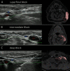
Ultrasound imaging at time of injection corresponding to acquired MRIs for each study group. Representative images of superficial (A), intermediate (B) and deep (C) cervical plexus blocks using ultrasound at time of injection (left) and MRI with contrast dye enhancement after injection (right). Blue line represent superficial fascia of the neck and green line represent deep fascia of the neck.CA, carotid artery; IJV, internal jugular vein; post. tub., posterior tubercle of the transverse process of the fourth cervical vertebrae.

References

    1. Greengrass RA, Narouze S, Bendtsen TF, et al. . Cervical plexus and greater occipital nerve blocks: controversies and technique update. Reg Anesth Pain Med 2019;44:623–6. 10.1136/rapm-2018-100261 - DOI - PubMed
    1. Koköfer A, Nawratil J, Opperer M. [Regional anesthesia for carotid surgery : An overview of anatomy, techniques and their clinical relevance]. Anaesthesist 2017. 10.1007/s00101-017-0270-z - DOI - PubMed
    1. Stoneham MD, Stamou D, Mason J. Regional anaesthesia for carotid endarterectomy. Br J Anaesth 2015;114:372–83. 10.1093/bja/aeu304 - DOI - PubMed
    1. Nash L, Nicholson HD, Zhang M. Does the investing layer of the deep cervical fascia exist? Anesthesiology 2005;103:962–8. 10.1097/00000542-200511000-00010 - DOI - PubMed
    1. Pandit JJ, Dutta D, Morris JF. Spread of injectate with superficial cervical plexus block in humans: an anatomical study. Br J Anaesth 2003;91:733–5. 10.1093/bja/aeg250 - DOI - PubMed

Publication types

LinkOut - more resources