. 2022 Jan 10;13(1):33.
doi: 10.1038/s41419-021-04424-x.

Microglial replacement in the aged brain restricts neuroinflammation following intracerebral hemorrhage

Affiliations

Microglial replacement in the aged brain restricts neuroinflammation following intracerebral hemorrhage

Xiuping Li et al. Cell Death Dis. .

Abstract

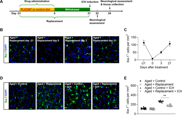
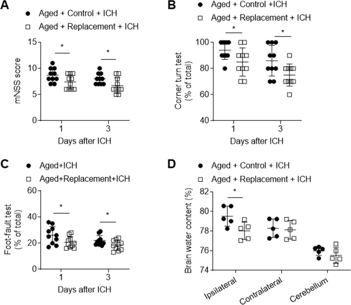
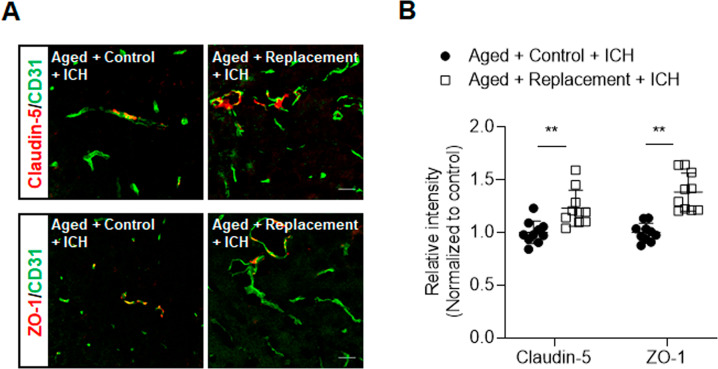
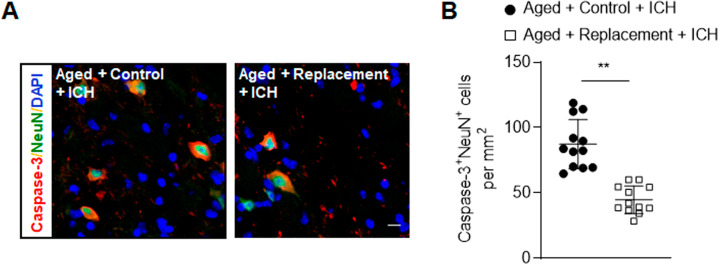
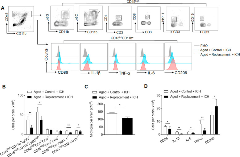
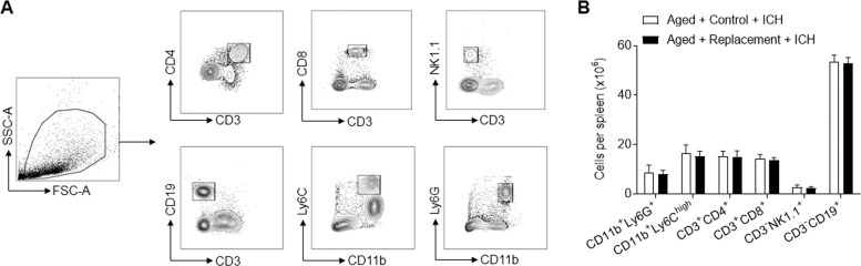
Aged microglia display augmented inflammatory activity after neural injury. Although aging is a risk factor for poor outcome after brain insults, the precise impact of aging-related alterations in microglia on neural injury remains poorly understood. Microglia can be eliminated via pharmacological inhibition of the colony-stimulating factor 1 receptor (CSF1R). Upon withdrawal of CSF1R inhibitors, microglia rapidly repopulate the entire brain, leading to replacement of the microglial compartment. In this study, we investigated the impact of microglial replacement in the aged brain on neural injury using a mouse model of intracerebral hemorrhage (ICH) induced by collagenase injection. We found that replacement of microglia in the aged brain reduced neurological deficits and brain edema after ICH. Microglial replacement-induced attenuation of ICH injury was accompanied with alleviated blood-brain barrier disruption and leukocyte infiltration. Notably, newly repopulated microglia had reduced expression of IL-1β, TNF-α and CD86, and upregulation of CD206 in response to ICH. Our findings suggest that replacement of microglia in the aged brain restricts neuroinflammation and brain injury following ICH.


Conflict of interest statement

The authors declare no competing interests.

Figures

Fig. 1
Fig. 1. Microglial replacement in the aged brain.
Groups of aged mice received 21 days of PLX3397 treatment in chow or a control diet, and then followed by three weeks of withdrawal prior to ICH induction. ICH induction, neurological assessment and tissue collection were conducted at indicated time points. A Flow chart illustrates drug administration and experimental design. B Brain sections from groups of aged mice receiving PLX3397 in chow at indicated time points after treatment or withdrawal were stained with Iba-1 (green). Nuclei were stained with DAPI (blue). Scale bar: 50 µm. C Counts of Iba-1+ cells in indicated groups of mice. n = 8 per group. D Brain sections from groups of aged mice receiving indicated treatments were stained with Iba-1 (green). Nuclei were stained with DAPI (blue). Scale bar: 50 µm. E Counts of Iba-1+ cells in indicated groups of mice. n = 12 per group. Data are presented as mean ± SD. **p < 0.01.
Fig. 2
Fig. 2. Effects of microglial replacement on neurological deficits and brain water content in the aged mice following ICH.
ICH was induced in mice by injection of collagenase. AC Summarized results showing neurological assessment (mNSS score, corner-turning test, and foot-fault test) of indicated groups of mice. n = 10 per group. D Brain water content in groups of mice receiving indicated treatments at day 3 after ICH. n = 5 per group. Data are presented as mean ± SD. *p < 0.05.
Fig. 3
Fig. 3. Effects of microglial replacement on blood-brain barrier integrity after ICH in the aged mice.
ICH was induced in mice by injection of collagenase. A Brain sections from ICH mice in microglial repopulation group or control group were stained with CD31 (green), Claudin-5 (red), and ZO-1 (red) at day 3 after ICH. Scale bar: 20 µm. B Summarized results show that microglial repopulation had reduced Claudin-5 and ZO-1 loss in immunofluorescence intensity within the lesion area after ICH in aged mice. n = 10 sections from two mice per group. Data are presented as mean ± SD. **p < 0.01.
Fig. 4
Fig. 4. Effects of microglial replacement on neuronal death after ICH in the aged mice.
ICH was induced in mice by injection of collagenase. A Brain sections from groups of ICH mice receiving indicated treatments after ICH were stained with NeuN (green) and Caspase-3 (red) at day 3 after ICH. Nuclei were stained with DAPI (blue). Scale bar: 10 µm. B Counts of Caspase-3+NeuN+ cells from groups of ICH mice receiving indicated treatments after ICH. n = 12 per group. Data are presented as mean ± SD. **p < 0.01.
Fig. 5
Fig. 5. Effects of microglial replacement on leukocyte infiltration and microglia activity in the aged brain following ICH.
ICH was induced in mice by injection of collagenase. At day 3 after ICH, brain tissues were obtained for flow cytometry analysis. A Gating strategy of brain-infiltrating neutrophils (CD45highCD11b+Ly6G+), monocytes (CD45highCD11b+Ly6Chigh), NK cells (CD45highCD3-NK1.1+), CD4+ T cells (CD45highCD3+CD4+), CD8+ T cells (CD45highCD3+CD8+), and B cells (CD45highCD3-CD19+), as well as microglia (CD11b+CD45int) and markers of their inflammatory activity (CD86, TNF-α, IL-6, IL-1β, CD206). B Counts of brain-infiltrating leukocyte subsets in the brains from indicated groups of ICH mice. C, D Counts of microglia and their expression CD86, TNF-α, IL-6, IL-1β, and CD206 in the brains from indicated groups of aged mice after ICH. n = 5 mice per group. Data are presented as mean ± SD. *p < 0.05, **p < 0.01.
Fig. 6
Fig. 6. Effects of microglial replacement on splenocytes in the aged mice following ICH.
ICH was induced in mice by injection of collagenase. At day 3 after ICH, spleen tissues were obtained for flow cytometry analysis. A Gating strategy of neutrophils (CD11b+Ly6G+), monocytes (CD11b+Ly6Chigh), NK cells (CD3-NK1.1+), CD4+ T cells (CD3+CD4+), CD8+ T cells (CD3+CD8+), and B cells (CD3-CD19+). B Cell counts of indicated immune cell subsets in spleens of aged mice after ICH. n = 5 per group. Data are presented as mean ± SD.

Similar articles

Cited by

References

    1. Perry VH, Holmes C. Microglial priming in neurodegenerative disease. Nat Rev Neurol. 2014;10:217–24. doi: 10.1038/nrneurol.2014.38. - DOI - PubMed
    1. Niraula A, Sheridan JF, Godbout JP. Microglia priming with aging and stress. Neuropsychopharmacology. 2017;42:318–33. doi: 10.1038/npp.2016.185. - DOI - PMC - PubMed
    1. Lan X, Han X, Li Q, Yang QW, Wang J. Modulators of microglial activation and polarization after intracerebral haemorrhage. Nat Rev Neurol. 2017;13:420–33. doi: 10.1038/nrneurol.2017.69. - DOI - PMC - PubMed
    1. Elmore MR, Najafi AR, Koike MA, Dagher NN, Spangenberg EE, Rice RA, et al. Colony-stimulating factor 1 receptor signaling is necessary for microglia viability, unmasking a microglia progenitor cell in the adult brain. Neuron. 2014;82:380–97. doi: 10.1016/j.neuron.2014.02.040. - DOI - PMC - PubMed
    1. Rice RA, Spangenberg EE, Yamate-Morgan H, Lee RJ, Arora RP, Hernandez MX, et al. Elimination of microglia improves functional outcomes following extensive neuronal loss in the hippocampus. J Neurosci. 2015;35:9977–89. doi: 10.1523/JNEUROSCI.0336-15.2015. - DOI - PMC - PubMed

Publication types

MeSH terms