. 2022 Jan 10;13(1):83.
doi: 10.1038/s41467-021-27648-z.

A mass spectrometric method for in-depth profiling of phosphoinositide regioisomers and their disease-associated regulation

Affiliations

A mass spectrometric method for in-depth profiling of phosphoinositide regioisomers and their disease-associated regulation

Shin Morioka et al. Nat Commun. .

Abstract

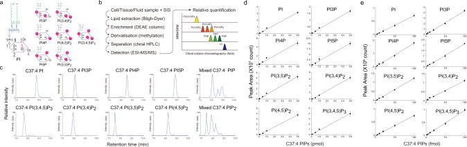
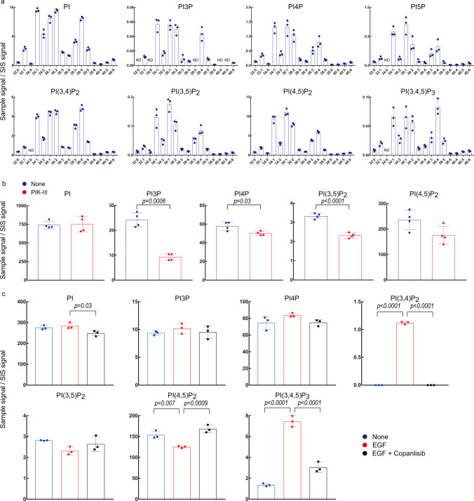
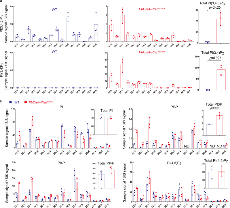
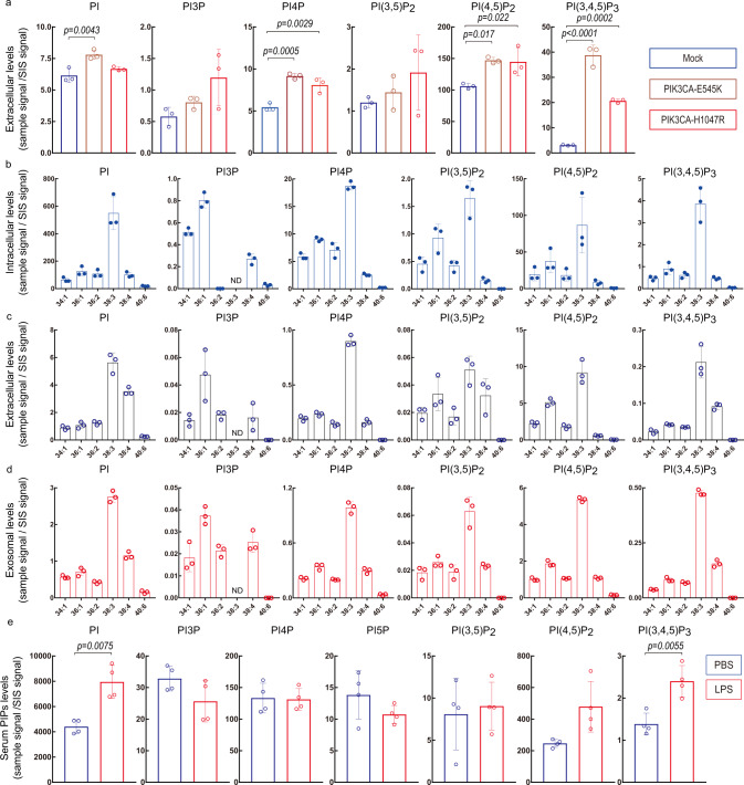
Phosphoinositides are a family of membrane lipids essential for many biological and pathological processes. Due to the existence of multiple phosphoinositide regioisomers and their low intracellular concentrations, profiling these lipids and linking a specific acyl variant to a change in biological state have been difficult. To enable the comprehensive analysis of phosphoinositide phosphorylation status and acyl chain identity, we develop PRMC-MS (Phosphoinositide Regioisomer Measurement by Chiral column chromatography and Mass Spectrometry). Using this method, we reveal a severe skewing in acyl chains in phosphoinositides in Pten-deficient prostate cancer tissues, extracellular mobilization of phosphoinositides upon expression of oncogenic PIK3CA, and a unique profile for exosomal phosphoinositides. Thus, our approach allows characterizing the dynamics of phosphoinositide acyl variants in intracellular and extracellular milieus.


Conflict of interest statement

H.N., J.S., and T.S. have filed for a patent (appl. No. 2018–528909), related to data in this paper on the phosphoinositide measurement method, which is under review. H.N serves as the CEO of and holds equity in LIPIDOME LAB Co., Ltd. T.S. is the founder of Japan Lipid Technologies, LLC. T.Y. serves as the CEO of Japan Lipid Technologies, LLC. The remaining authors declare no competing interests.

Figures

Fig. 1
Fig. 1. Phosphoinositide regioisomer measurement by PRMC-MS.
a Schematic structures of the eight phosphoinositide classes. Note that phosphatidylinositol mono- and bisphosphate (PIP and PIP2) classes each contain three regioisomers. b Scheme depicting the general workflow of PRMC-MS. SIS, surrogate internal standard lipids. c Mass spectra from PRMC-MS analyses of C37:4 (1-heptadecanoyl-2-eicosatetraenoyl) standard chemicals representing each of the eight phosphoinositide classes. Mixed, a mixture of PIP or PIP2 regioisomers. d, e Linear regression analyses of peak area vs pmol (d) and fmol (e) concentrations of the indicated phosphoinositides. Amounts of standard phosphoinositides analyzed were 0.002, 0.005, 0.01, 0.02, 0.05, 0.1, 0.2, 0.5, 1, 5, 10, 50 pmol in d and 2, 5, 10, 20, 50, 100 fmol in e (see also Supplementary Table 1). The C37:4 (1-heptadecanoyl-2-eicosatetraenoyl) molecular species of each phosphoinositide class was used. Data were the mean ± SD (n = 4 technical replicates). Source data are provided as a Source Data file.
Fig. 2
Fig. 2. Application of PRMC-MS to phosphoinositide measurement in cell lines.
a Levels of the indicated phosphoinositide acyl variants in HEK293T cells. PI, PI3P, PI4P, PI(3,5)P2, PI(4,5)P2, and PI(3,4,5)P3 were assessed in mock-transfected cells. PI5P was assessed in cells overexpressing IpgD. PI(3,4)P2 was assessed in non-transfected cells treated with 1 mM H2O2 for 5 min. Data were the mean ± SD (n = 3 biologically independent samples). ND not determined. b Phosphoinositide measurements in HeLa cells treated with vehicle (control) or 5 µM PIK-III for 2 h. PI5P, PI(3,4)P2, and PI(3,4,5)P3 were not detected. Data were the mean ± SD of total acyl variants of the indicated phosphoinositide classes (n = 4 biologically independent samples), and p values by two-sided Welch’s t-test are shown. c Phosphoinositide measurements in PC3 cells that were left untreated (control), or treated with vehicle or 100 nM copanlisib for 30 min prior to stimulation with 10 ng/ml EGF. PI5P was not detected. Data were the mean ± SD of total acyl variants of the indicated phosphoinositide classes (n = 3 biologically independent samples). The significance was analyzed using a two-sided Dunnett’s t-test. Source data are provided as a Source Data file.
Fig. 3
Fig. 3. Application of PRMC-MS to phosphoinositide measurement in mouse tissue.
a Left and middle panels: Quantification of the indicated PI(3,4,5)P3 and PI(3,4)P2 acyl variants in prostate tissues from WT and prostate-specific Pten-deficient (PbCre4-Ptenflox/flox) mice at 12 weeks of age (n = 3 independent animals/group). The altered acyl chain profile in Pten-deficient cancerous tissues was confirmed in five independent experiments. Right panel: Total of the PI(3,4)P2 and PI(3,4,5)P3 acyl variants in the tissues in the left and middle panels. b Measurements of the indicated acyl variants and total PI, PI3P, PI4P, and PI(4,5)P2 in the prostate tissues from the WT (blue) and PbCre4-Ptenflox/flox (red) mice in (a) (12 weeks of age, n = 3). For all panels, data were the mean ± SD (n = 3 independent animals). ND not determined. The significance between WT and PbCre4-Ptenflox/flox mice was analyzed using a two-sided Welch’s t-test. Source data are provided as a Source Data file.
Fig. 4
Fig. 4. Application of PRMC-MS to phosphoinositide measurement in liquid samples.
a Quantification of the indicated phosphoinositides in CM of HEK293T cells transfected with either control expression vector (Mock) or vector expressing either mutant E545K or H1047R PIK3CA. CM was collected at 24 h post-transfection. Data were the mean ± SD (n = 3 biologically independent samples) and representative of three independent experiments. Statistical analysis was performed using a two-sided Dunnett’s t-test. bd Quantification of the indicated phosphoinositide acyl variants in b cell pellets, c CM, and d exosomes derived from LNCap cell cultures. PI5P and PI(3,4)P2 were not detected. Data were the mean ± SD (n = 3 biologically independent samples). e Quantification of the indicated phosphoinositides in plasma from WT mice that were intraperitoneally injected with either vehicle (PBS) or 50 mg/ml lipopolysaccharide (LPS) and analyzed after 2 h. PI(3,4)P2 was not detected in either case. Data were the mean ± SD of total acyl variants of the indicated phosphoinositide classes (n = 4 biologically independent samples). Statistical analysis was performed using two-sided unpaired Welch’s t-test. Source data are provided as a Source Data file.

References

    1. Balla T. Phosphoinositides: tiny lipids with giant impact on cell regulation. Physiol. Rev. 2013;93:1019–1137. doi: 10.1152/physrev.00028.2012. - DOI - PMC - PubMed
    1. Sasaki T, et al. Mammalian phosphoinositide kinases and phosphatases. Prog. Lipid Res. 2009;48:307–343. doi: 10.1016/j.plipres.2009.06.001. - DOI - PubMed
    1. Hammond GR, Balla T. Polyphosphoinositide binding domains: key to inositol lipid biology. Biochim. Biophys. Acta. 2015;1851:746–758. doi: 10.1016/j.bbalip.2015.02.013. - DOI - PMC - PubMed
    1. Goncalves MD, Hopkins BD, Cantley LC. Phosphatidylinositol 3-kinase, growth disorders, and cancer. N. Engl. J. Med. 2018;379:2052–2062. doi: 10.1056/NEJMra1704560. - DOI - PubMed
    1. Fruman DA, et al. The PI3K pathway in human disease. Cell. 2017;170:605–635. doi: 10.1016/j.cell.2017.07.029. - DOI - PMC - PubMed

Publication types

MeSH terms