. 2022 Jan 10;13(1):66.
doi: 10.1038/s41467-021-27650-5.

TASOR epigenetic repressor cooperates with a CNOT1 RNA degradation pathway to repress HIV

Affiliations

TASOR epigenetic repressor cooperates with a CNOT1 RNA degradation pathway to repress HIV

Roy Matkovic et al. Nat Commun. .

Abstract

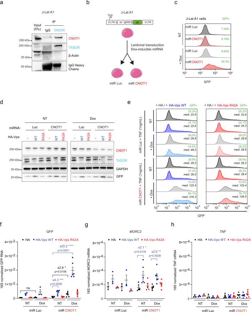
The Human Silencing Hub (HUSH) complex constituted of TASOR, MPP8 and Periphilin recruits the histone methyl-transferase SETDB1 to spread H3K9me3 repressive marks across genes and transgenes in an integration site-dependent manner. The deposition of these repressive marks leads to heterochromatin formation and inhibits gene expression, but the underlying mechanism is not fully understood. Here, we show that TASOR silencing or HIV-2 Vpx expression, which induces TASOR degradation, increases the accumulation of transcripts derived from the HIV-1 LTR promoter at a post-transcriptional level. Furthermore, using a yeast 2-hybrid screen, we identify new TASOR partners involved in RNA metabolism including the RNA deadenylase CCR4-NOT complex scaffold CNOT1. TASOR and CNOT1 synergistically repress HIV expression from its LTR. Similar to the RNA-induced transcriptional silencing complex found in fission yeast, we show that TASOR interacts with the RNA exosome and RNA Polymerase II, predominantly under its elongating state. Finally, we show that TASOR facilitates the association of RNA degradation proteins with RNA polymerase II and is detected at transcriptional centers. Altogether, we propose that HUSH operates at the transcriptional and post-transcriptional levels to repress HIV proviral expression.


Conflict of interest statement

The authors declare no competing interests.

Figures

Fig. 1
Fig. 1. TASOR destabilizes HIV-1 LTR-driven transcripts.
a HeLa HIV-1 LTR-ΔTAR-Luc cell model. HeLa HIV-1 LTR-Luc and HeLa HIV-1 LTRΔTAR-Luc cells were lysed for Luc RNA quantification (n = 3; each replicate is presented along with the mean values and the SEM. A two-sided unpaired t test was used. ****p < 0.0001). b TASOR overexpression decreases LTR-driven Luc expression. HeLa HIV-1 LTR-ΔTAR-Luc were TASOR-depleted by transient transfection of the pLenti-CRISPR-V2 and a sgRNA guide targeting the first exon of TASOR. These TASOR-depleted cells were then transfected with increasing amounts of HA-TASOR WT or HA-TASORΔPARP encoding pAS1B vectors. Luc activity was measured and proteins were analyzed by western blot 48 h post-transfection (n = 3; each independent replicate is presented along with the mean values and the SEM. A two-sided unpaired t test was used. **p = 0.0029; *p = 0.0262. c TASOR negatively impacts LTR-driven Luc transcript at a post-transcriptional step. HeLa HIV-1 LTRΔTAR-Luc were transfected for 72 h with siCtrl or siTASOR. The luciferase activity was measured and Nuclear Run On experiments were performed (n = 3, each independent replicate is presented along with the mean values and the SEM). d HIV-2 Vpx mimics siRNA-mediated TASOR silencing in increasing LTR-driven transcript stability. Nuclear Run On performed in HeLa HIV-1 LTRΔTAR-Luc after 48 h of pAS1B-HA, pAS1B-HA-Vpx WT or R42A HIV-2 Ghana, and pAS1B-HA-Vpr HIV-1 transfection. The HIV-1 LTR-driven luciferase RNA expression was measured by RT-qPCR (n = 4, each independent replicate is presented along with the mean values and the SEM) and a western blot analysis monitored the levels of expression of the lentiviral proteins, TASOR, and HLTF (as an HIV-1 Vpr target). Source data are provided as a Source data file.
Fig. 2
Fig. 2. TASOR interacts and cooperates with the CCR4-NOT complex scaffold CNOT1 to destabilize LTR-driven transcripts.
a Identification of CNOT1 and other proteins as new partners of TASOR by yeast-two-hybrid screening. Proteins involved in transcriptional regulation, in post-transcriptional regulation of RNAs, and in mRNA degradation pathways are colored in red, blue, and green, respectively. Newly found interactions are shown in blue. Those validated by at least 3 coIPs are shown in solid line (TASOR-CNOT1 validated in Figs. 2b–e, 3a, 6b–d, 7g, and S3c, TASOR-SUPTH6 in Fig. S2g, TASOR-MATRIN 3 in Fig. 6d, TASOR-ZFC3H1 in Fig. S3c). b TASOR interacts with CNOT1 by co-immunoprecipitation. Mock, DDK, and TASOR-DDK vectors were transfected in HeLa cells and anti-DDK immunoprecipitation was performed (n > 3, some are presented all along this study). c Endogenous TASOR interacts with endogenous CNOT1 in the nucleus of HeLa HIV-1 LTR-ΔTAR-Luc cells. GAPDH is a negative control. (n = 2); d a transcript stabilizes TASOR-CNOT1 interaction. DDK and TASOR-DDK vectors were transfected in HeLa cells. Lysates were treated or not with DNase 1 or RNase A. Anti-DDK immunoprecipitation was then performed. IGF2BP1 is a negative control (n > 3). e The PARP domain of TASOR is required for its interaction with CNOT1. HeLa HIV-1 LTR-ΔTAR-Luc cells were transfected with pLenti-TASOR WT-DDK or TASORΔPARP-DDK vectors. An anti-DDK IP and a western blot analysis were performed to assess the interactions with CNOT1 (n > 3). f TASOR and CNOT1 cooperate to repress HIV-1 LTR expression at a post-transcriptional level. After siRNA transfection in HeLa HIV-1 LTR-ΔTAR-Luc, a Nuclear Run On experiment was undertaken to measure LTR-driven Luc transcripts levels. (n = 3; each color represents one different independent experiment, mean and SEM are shown). g TASOR represses MORC2 transcription while CNOT1 decreases its stability. After siRNA transfection in HeLa HIV-1 LTR-ΔTAR-Luc, a Nuclear Run On experiment was undertaken to measure MORC2 transcripts levels (n = 3; each color represents one different independent experiment, mean and SEM are shown). h CNOT1 silencing only increases TNFα transcript stability. After siRNA transfection in HeLa HIV-1 LTR-ΔTAR-Luc, a Nuclear Run On experiment was undertaken to measure TNFα transcripts levels (n = 3; each color represents one different independent experiment, mean and SEM are shown). Source data are provided as a Source data file.
Fig. 3
Fig. 3. TASOR and CNOT1 cooperate to repress HIV-1 expression in the J-Lat A1 model of HIV-1 latency.
a TASOR interacts with CNOT1 in the J-Lat A1 T cell line. TASOR or Rabbit IgG immunoprecipitations were performed in J-Lat A1 lysates. The interaction was then assessed by western blot. β-Actin is a negative control. (n = 2 in Jurkat cell lines). b Generation of Dox-inducible miR-RNA CNOT1 J-Lat A1 cells. J-Lat A1 cells, harboring one copy of integrated and latent LTR-tat-IRES-GFP-LTR construct, were transduced with a pINDUCER10 vector containing a doxycycline-inducible miRNA targeting CNOT1 or Luciferase (Mock) transcripts. c Expression of the miRNA targeting CNOT1 reactivates GFP expression from the latent HIV-1 minigenome. J-Lat A1 miR Luc or miR CNOT1 were treated or not with doxycyline 1 µg/mL for 72 h and GFP expression was analyzed by flow cytometry. The proportion of cells that became GFP positive is indicated in green. d Dox-inducible miR-CNOT1, but not miR-Luc, decreases CNOT1 expression while delivery of VLPs containing HIV-2 Vpx WT induces TASOR depletion in contrast to Vpx R42A in the J-Lat A1 cells. J-Lat A1 miR Luc and miR CNOT1 were treated or not for 72 h with doxycycline 1 µg/mL and VLPs containing HA-Vpx WT or R42A or mock were delivered for 24 h prior to protein analysis. ef Co-silencing of CNOT1 and TASOR synergistically increases LTR-driven GFP expression at the protein level with the proportion of cells that became GFP positive indicated in green and the median of GFP expression in the whole cell population in italic (e), at the RNA level (f), but not MORC2 RNA (g) or TNF RNA (h) in the J-Lat A1 model. J-Lat A1 miR Luc and miR CNOT1 were treated or not for 72 h with doxycycline 1 µg/mL and VLPs containing HA-Vpx WT or R42A or mock were delivered for 24 h and TNF (1 ng/mL) 16 h prior to flow cytometric analysis and RT-qPCR analyses (from d to h, n = 3 for miR Luc; n = 6 for miR CNOT1; each independent replicate, mean and SEM are shown; two-sided unpaired t test was applied). Source data are provided as a Source data file.
Fig. 4
Fig. 4. TASOR and CNOT1 cooperate to repress HIV-1 expression in latently HIV-1-infected Jurkat T cells.
a Schematic of the LTR-EGFP provirus used to analyze HIV-1 LTR-driven EGFP expression in infected Jurkat T cells. Primers used to quantify LTR-driven transcripts are mapped in orange. b Generation of latently HIV-1-infected Jurkat cells based on the EGFP expression by flow cytometry. The sorted cells were then transduced with the doxycycline-inducible miR CNOT1 or Luc vectors. Without doxycycline treatment, cells remained EGFP negative by flow cytometry, whereas LTR-driven RNAs (US) were detected by RT-qPCR in the latently infected cells. RNA was normalized on the 18s rRNA (n = 2). c Dox-inducible miR-CNOT1, but not miR-Luc, decreases CNOT1 expression while the delivery of VLPs containing HIV-2 Vpx WT induces TASOR depletion in contrast to Vpx R42A in these latently infected Jurkat cells. The latently HIV-1-infected Jurkat miR Luc and miR CNOT1 cells were treated or not for 72 h with doxycycline 1 µg/mL, with VLPs containing HA-Vpx WT or R42A or mock were delivered for 24 h and with TNF (1 ng/mL) 16 h prior to protein analysis. The co-depletion of CNOT1 and TASOR synergistically increases HIV-1 LTR at the protein level with the proportion of cells that became EGFP positive indicated in green and the median of GFP expression in the whole cell population in italic (d), at the RNA level (e), but not MORC2 RNA (f) or TNF RNA (g) in these latently HIV-1-infected T cell lines. The latently HIV-1-infected Jurkat miR Luc and miR CNOT1 cells were treated or not for 72 h with doxycycline 1 µg/mL and VLPs containing HA-Vpx WT or R42A or mock were delivered for 24 h and TNF (1 ng/mL) 16 h prior to flow cytometric analysis and RT-qPCR analyses (for dg, n = 3 independent experiments, mean and SEM are represented; two-sided unpaired t test was applied: p values are indicated in the graph). Source data are provided as a Source data file.
Fig. 5
Fig. 5. TASOR cooperates with CNOT1 to repress a subset of genes in HeLa cells.
a HeLa cells were transfected for 72 h with siRNA targeting TASOR or CNOT1 or both transcripts. A western blot assessed the downregulation of TASOR and CNOT1 prior to RNAseq analyses. (western blot of the three different replicates is presented). b Number of genes that are significantly upregulated (by at least 20%), upon TASOR + CNOT1 co-silencing as compared to control, TASOR, or CNOT1 individual silencing in HeLa cells (log2 fold change > 0.2630344; p adjusted values < 0.05 after Benjamini–Hochberg multiple testing correction of Wald test p value). c Cooperativity of TASOR and CNOT1 regulation on host genes. The interaction term of the model used for differential gene expression analysis (∆contrast, DESeq2). d Cooperative effects affecting genes upregulated in one or the other of the single siRNA conditions (siTASOR or siCNOT1) and not downregulated in the second condition (log2 fold change > 0; p adjusted values < 0.05 after Benjamini–Hochberg multiple testing correction of Wald test p value). Fold changes (FC1 and FC2, as defined above) for individual genes were plotted showing the potential interaction modes of TASOR and CNOT1. e MA plot representing the differential expression of transposable element (TE) families in HeLa cells, upon co-silencing of TASOR and CNOT1 as compared to cells transfected with control siRNA. Significantly upregulated and downregulated TE families are highlighted in red and blue, respectively (log2(FC) > 0.2630344; p adjusted values < 0.05, Wald test with Benjamini–Hochberg multiple testing correction). ns not significant. f Number of TE families that are significantly upregulated (by at least 20%), upon TASOR + CNOT1 co-silencing as compared to control, TASOR, or CNOT1 individual silencing in HeLa cells (log2 fold change > 0.2630344; p adjusted values < 0.05 after Benjamini–Hochberg multiple testing correction of Wald test p value). More information is available in Source data file.
Fig. 6
Fig. 6. TASOR interacts and cooperates with nuclear RNA degradation factors.
a TASOR is a nuclear protein. Cytoplasmic and nuclear protein extracts from HeLa HIV-1 LTR-ΔTAR-Luc cells were loaded on a SDS-PAGE gel. GAPDH and MATR3 are markers of the cytoplasmic and nuclear fractions respectively (n > 3). b Endogenous TASOR interacts with the endogenous, nuclear CNOT1, and its partners CNOT7, YTHDF2, the endogenous TRAMP-like/NEXT/PAXT component MTR4, and the endogenous RNA exosome factor EXOSC10. HeLa HIV-1 LTR-ΔTAR-Luc cells were fractionated and endogenous TASOR immunoprecipitation was performed in the nuclear fraction. Lamin B1 is negative control (n > 3). c Reverse immunoprecipitation confirmed the interaction between endogenous CNOT1 and MTR4 proteins with endogenous TASOR in the nucleus. HeLa HIV-1 LTR-ΔTAR-Luc cells were fractionated and endogenous CNOT1 and MTR4 immunoprecipitation was performed in the nuclear fraction. The asterisk shows the SUPT6H band (n = 3). d TASOR interacts with the known CNOT1 partners in an RNA-dependent manner. DDK and TASOR-DDK vectors were transfected in HeLa HIV-1 LTR-ΔTAR-Luc cells. After 48 h, lysates were treated or not with RNase A for 30 min at room temperature. Anti-DDK immunoprecipitation was then performed. All lanes are from the same gel (n > 3). e TASOR cooperates with the nuclear RNA destabilization/degradation factors. After 72 h of siRNA transfections in HeLa HIV-1 LTR-ΔTAR-Luc, cells were lysed and luciferase activity was measured and normalized on protein concentration (n = 5 for the siRNA TASOR and siRNA CNOT1 conditions; n = 3 for other siRNA transfections; each color represents one different independent experiment, mean and SEM are shown). f Schematic representation of RNA-mediated interactions between TASOR and CNOT1 and its partners: the TRAMP-like/NEXT/PAXT component MTR4, the Nuclear Exosome, and the m6A reader YTHDF2. Source data are provided as a Source data file.
Fig. 7
Fig. 7. TASOR recruits RNA-degradation factors onto elongating RNAPII to silence gene expression.
a TASOR colocalizes with active HIV-1 transcriptional centers in HIV-1-infected Jurkat cells. Latently HIV-1-EGFP-infected Jurkat cells were transduced with VLPs containing Vpx R42A or WT for 24 h and treated with TNF (1 ng/mL). LTR-driven unspliced (us) RNAs were marked with egfp probes and TASOR by immunofluorescence. The graph corresponds to the signal intensities across the white line. b Vpx WT triggers a significant loss of TASOR at the HIV-1 transcriptional centers. TASOR signal at HIV-1 transcription centers in the Vpx R42A delivery condition was set as 100%. (n = 261, n = 267, and n = 66 for the Vpx R42A, Vpx WT, and secondary antibody (2nd.ab) Alexa 594 conditions, respectively; two-sided unpaired t test was applied: ****p < 0.001). c TASOR interacts with endogenous RNAPII in HeLa HIV-1 LTR-ΔTAR-Luc cells. GAPDH is a negative control (n > 3). d TASOR affinity for RNAPII is greatly reduced upon depletion of the N-terminal PARP domain. Lamin B1 and GAPDH are negative controls (n = 3). e TASOR interacts with elongating RNAPII in HEK293T and HeLa HIV-1 LTR-ΔTAR-Luc cells. Ser5-phosphorylated and Ser2-phosphorylated RNAPII are markers of RNAPII in the initiating and elongating phases of transcription, respectively, MPP8 is a positive control. GAPDH is a negative control (n > 3). f TASOR recruits CNOT1 and its partners onto RNAPII. DDK and TASOR-DDK vectors were transfected in HeLa HIV-1 LTR-ΔTAR-Luc cells. TASOR was revealed either with an anti-DDK (TASOR-DDK) or with an anti-TASOR to detect the endogenous protein. SUPT6H is a control of a known RNAPII partner. MORC2 is necessary for HUSH-mediated gene silencing according to ref. . GAPDH is a negative control (n = 3). g TASOR and CNOT1 are found in complex with the HIV-1 nascent transcript. Nascent RNAs from HeLa HIV-1 LTR-ΔTAR-Luc cells were labeled and anti-TASOR and anti-CNOT1 immunoprecipitations were performed from the nuclear extract. GAPDH and TASOR partner MATR3 are markers of the cytoplasmic and nuclear fractions, respectively. Immunoprecipitated RNAs were normalized on input signal (n = 4; mean and SEM are shown; two-sided unpaired t test was applied: purple stars or NS indicate statistical differences between TASOR and CNOT1 conditions; blue stars: TASOR vs IgG, red stars: CNOT1 vs IgG). Source data are provided as a Source data file.

References

    1. Jordan A, Defechereux P, Verdin E. The site of HIV-1 integration in the human genome determines basal transcriptional activity and response to Tat transactivation. EMBO J. 2001;20:1726–1738. - PMC - PubMed
    1. Lewinski MK, et al. Genome-wide analysis of chromosomal features repressing human immunodeficiency virus transcription. J. Virol. 2005;79:6610–6619. - PMC - PubMed
    1. Brady T, et al. HIV integration site distributions in resting and activated CD4+ T cells infected in culture. AIDS. 2009;23:1461–1471. - PMC - PubMed
    1. Marcello A, Dhir S, Dieudonne M. Nuclear positional control of HIV transcription in 4D. Nucleus. 2010;1:8–11. - PMC - PubMed
    1. Chougui G, et al. HIV-2/SIV viral protein X counteracts HUSH repressor complex. Nat. Microbiol. 2018;3:891–897. - PubMed

Publication types

MeSH terms