. 2022 Jan 10;13(1):80.
doi: 10.1038/s41467-021-27674-x.

Cross-reactive memory T cells associate with protection against SARS-CoV-2 infection in COVID-19 contacts

Affiliations

Cross-reactive memory T cells associate with protection against SARS-CoV-2 infection in COVID-19 contacts

Rhia Kundu et al. Nat Commun. .

Abstract

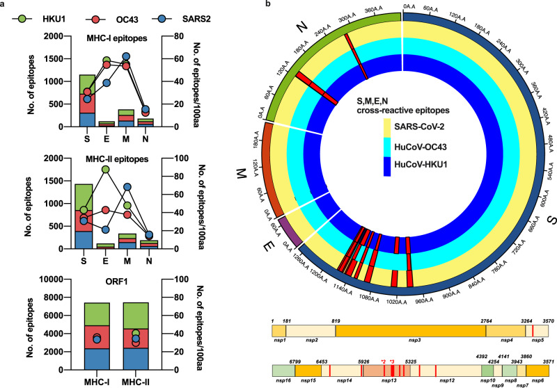
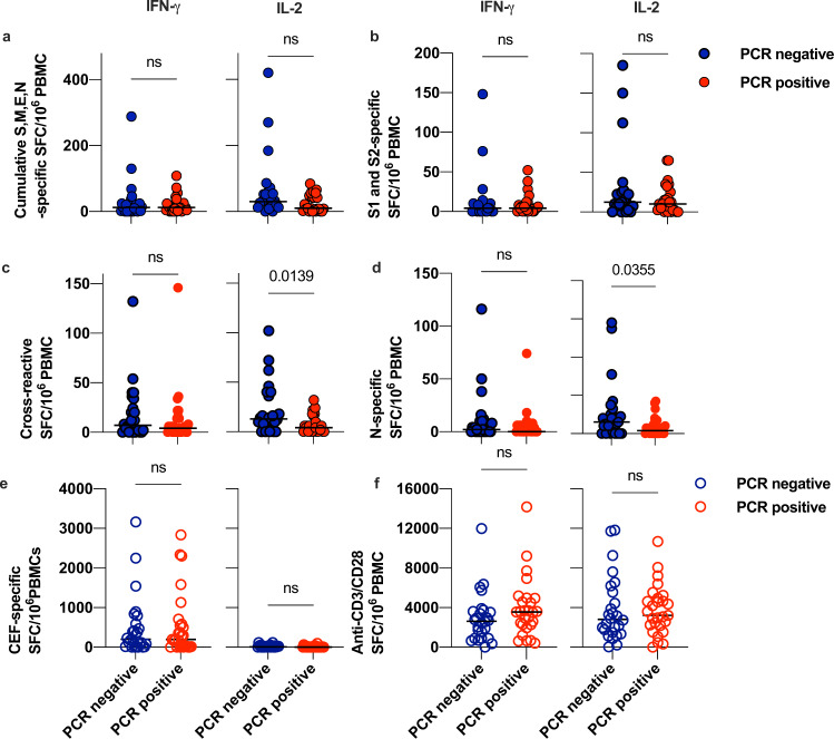
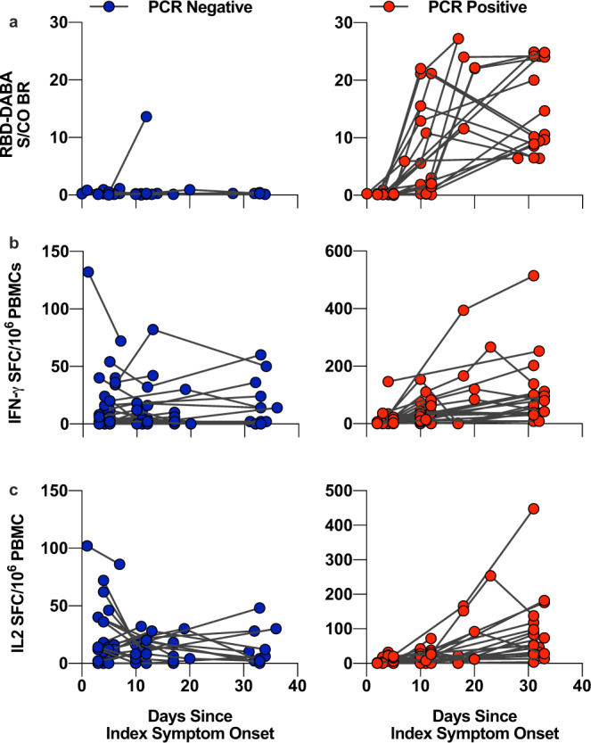
Cross-reactive immune responses to SARS-CoV-2 have been observed in pre-pandemic cohorts and proposed to contribute to host protection. Here we assess 52 COVID-19 household contacts to capture immune responses at the earliest timepoints after SARS-CoV-2 exposure. Using a dual cytokine FLISpot assay on peripheral blood mononuclear cells, we enumerate the frequency of T cells specific for spike, nucleocapsid, membrane, envelope and ORF1 SARS-CoV-2 epitopes that cross-react with human endemic coronaviruses. We observe higher frequencies of cross-reactive (p = 0.0139), and nucleocapsid-specific (p = 0.0355) IL-2-secreting memory T cells in contacts who remained PCR-negative despite exposure (n = 26), when compared with those who convert to PCR-positive (n = 26); no significant difference in the frequency of responses to spike is observed, hinting at a limited protective function of spike-cross-reactive T cells. Our results are thus consistent with pre-existing non-spike cross-reactive memory T cells protecting SARS-CoV-2-naïve contacts from infection, thereby supporting the inclusion of non-spike antigens in second-generation vaccines.


Conflict of interest statement

The authors declare no competing interests.

Figures

Fig. 1
Fig. 1. In silico prediction of putative epitopes in human endemic beta-coronaviruses and SARS-CoV-2.
Available sequences for S, M, E, N and ORF1 from SARS-CoV-2, huCoV-OC43 and huCoV-HKU1 were assessed for potential HLA-binding motifs by SYFPEITHI and IEDB as described in the Methods. The number of predicted epitopes is presented in the stacked bar charts and the immunogenicity for each protein of each virus is depicted as points on the right Y-axis. Green bars and circles depict huCoV-HKU1 (HKU1), red huCoV-OC43 (OC43) and blue SARS-CoV-2 (SARS2) (a). The predicted cross-reactive epitopes and their prevalence within each virus for all sequences available are mapped as red histograms in a linear plot for ORF1 and a radial plot for S, M, E and N. All mapped cross-reactive epitopes within ORF1 were present across SARS-CoV-2, HKU1 and OC43. Within the radial plot, the yellow track represents SARS-CoV-2 sequence, dark blue HKU1 and light blue OC43, with red histograms representing the prevalence of the putative epitope across the viruses (b).
Fig. 2
Fig. 2. Dual cytokine FLISpot responses to SARS-CoV protein-spanning peptide pools and the cross-reactive pool in PCR-positive and PCR-negative COVID-19 contacts.
PBMCs sampled from COVID-19 contacts (n = 52) at the baseline visit were rested overnight at high density prior to stimulation with 1 µg/ml/peptide pools, 1 µg/ml CMV-EBV-Flu (CEF) positive control peptide pool or 1 µg/ml anti-CD3 and 1 µg/ml anti-CD28 antibody as indicated and cultured for 20 h in a FLISpot assay to detect IL-2- (left-hand panels) and IFNγ (right-hand panels) secreting T cells. Data are DMSO-subtracted spot forming cells per 1 × 106 PBMC. Blue circles represent PCR-negative contacts and red circles represent PCR-positive contacts. Filled circles represent test data from SARS-CoV-2 stimuli and the outline circles represent values from positive control stimuli. a Depicts cumulative frequency of pan-S,M,E and N-specific T cells using protein-spanning 15-mer peptide pools to represent the entire protein, rather than predicted epitopes, as a stimuli. b Depicts the frequency of pan-S-specific T cells, using protein-spanning 15-mer peptide pools to represent the entire protein, rather than predicted epitopes, as stimuli. c Depicts the frequency of cross-reactive T cells, using peptides derived from the putative MHC-I and MHC-II epitopes defined in our bioinformatic analysis and Nelde et al, as listed in Supplementary Table 1. d Depicts the frequency of pan-N-specific T cells using protein-spanning 15-mer peptide pools to represent the entire protein, rather than predicted epitopes, as a stimuli. e Depicts the frequency of T cells specific to an antigen-specific positive control peptide pool comprising of well-characterised epitopes from influenza, cytomegalovirus and Epstein–Barr Virus. f Depicts the frequency of IL-2 and IFNγ secreting T cells activated in response to a polyclonal stimulus of anti-CD3 and anti-CD28 soluble antibody. Data are from n = 26 PCR-positive and n = 26 PCR-negative contacts and p-values are from a two-sided Welch’s T-test.
Fig. 3
Fig. 3. Dynamics of cross-reactive T cells and RBD-specific antibody in PCR-positive and negative contacts.
Serum sampled from COVID-19 contacts at the baseline, D7 and D28 visit were assayed for RBD-specific antibody, represented as sample/control ratios (a). PBMCs from these visits were rested overnight at high density prior to stimulation with 1 µg/ml cross-reactive peptide pool cultured for 20 h in a FLISpot assay to detect IL-2- (b) and IFNγ (c) secreting T cells. Serum from these visits were assayed for RBD-specific antibody, represented as sample/control ratios (c). Left-hand panels and blue circles represent PCR-negative contacts whilst right-hand panels and red circle represent PCR-positive contacts.

Comment in

  • Understanding Kids and COVID.
    Peeples L. Peeples L. Proc Natl Acad Sci U S A. 2022 Mar 29;119(13):e2203753119. doi: 10.1073/pnas.2203753119. Epub 2022 Mar 22. Proc Natl Acad Sci U S A. 2022. PMID: 35316141 Free PMC article. No abstract available.

References

    1. Mateus J, et al. Selective and cross-reactive SARS-CoV-2 T cell epitopes in unexposed humans. Science. 2020;370:89–94. doi: 10.1126/science.abd3871. - DOI - PMC - PubMed
    1. Grifoni A, et al. Targets of T cell responses to SARS-CoV-2 coronavirus in humans with COVID-19 disease and unexposed individuals. Cell. 2020;181:1489–1501.e15. doi: 10.1016/j.cell.2020.05.015. - DOI - PMC - PubMed
    1. Nelde, A. et al. SARS-CoV-2-derived peptides define heterologous and COVID-19-induced T cell recognition. Nat. Immunol. 10.1038/s41590-020-00808-x. - PubMed
    1. Le Bert N, et al. SARS-CoV-2-specific T cell immunity in cases of COVID-19 and SARS, and uninfected controls. Nature. 2020;584:457–462. doi: 10.1038/s41586-020-2550-z. - DOI - PubMed
    1. Ferretti AP, et al. Unbiased Screens Show CD8+ T Cells of COVID-19 Patients Recognize Shared Epitopes in SARS-CoV-2 that Largely Reside outside the Spike Protein. Immunity. 2020;53:1095–1107.e3. doi: 10.1016/j.immuni.2020.10.006. - DOI - PMC - PubMed

Publication types

MeSH terms