. 2022 Jan 10;8(1):7.
doi: 10.1038/s41420-021-00799-0.

Microfluidic deep mutational scanning of the human executioner caspases reveals differences in structure and regulation

Affiliations

Microfluidic deep mutational scanning of the human executioner caspases reveals differences in structure and regulation

Hridindu Roychowdhury et al. Cell Death Discov. .

Erratum in

Abstract

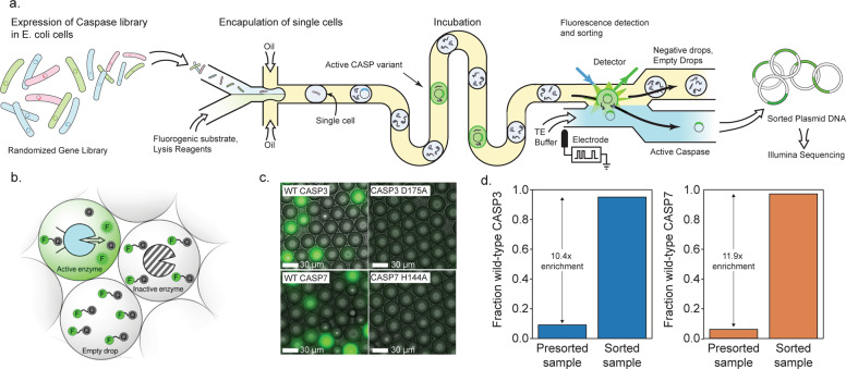
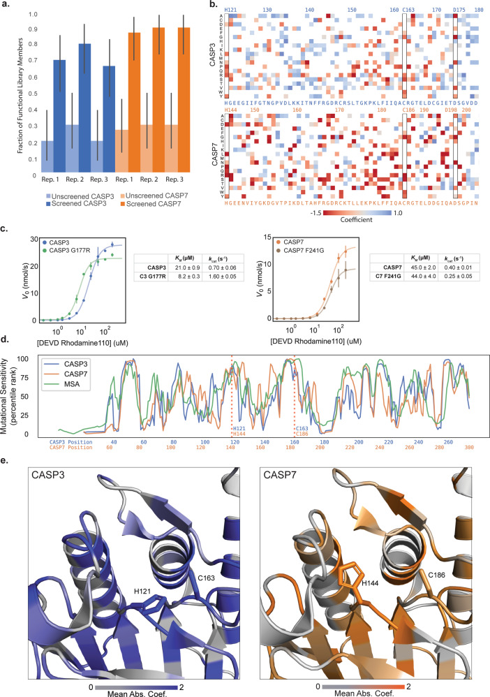
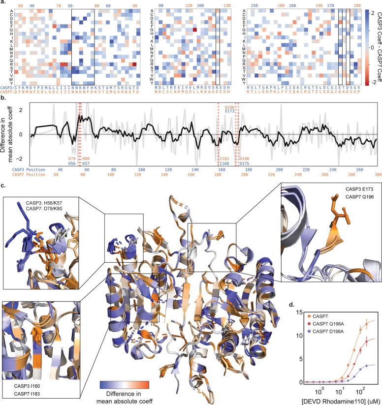
The human caspase family comprises 12 cysteine proteases that are centrally involved in cell death and inflammation responses. The members of this family have conserved sequences and structures, highly similar enzymatic activities and substrate preferences, and overlapping physiological roles. In this paper, we present a deep mutational scan of the executioner caspases CASP3 and CASP7 to dissect differences in their structure, function, and regulation. Our approach leverages high-throughput microfluidic screening to analyze hundreds of thousands of caspase variants in tightly controlled in vitro reactions. The resulting data provides a large-scale and unbiased view of the impact of amino acid substitutions on the proteolytic activity of CASP3 and CASP7. We use this data to pinpoint key functional differences between CASP3 and CASP7, including a secondary internal cleavage site, CASP7 Q196 that is not present in CASP3. Our results will open avenues for inquiry in caspase function and regulation that could potentially inform the development of future caspase-specific therapeutics.


Conflict of interest statement

The authors declare no competing interests.

Figures

Fig. 1
Fig. 1. A droplet microfluidic platform for screening caspases.
a A schematic of our microfluidic screening system. A dilute suspension of E. coli expressing caspase variants are injected into a microfluidic device and individual cells are encapsulated into microdroplets containing lysis reagents and a fluorogenic caspase substrate. The cells are lysed, the enzyme reaction is incubated on-chip, and the fluorescence of each droplet is analyzed using a laser. The fluorescent droplets are then sorted by electrocoalescence with an aqueous stream that collects the sorted plasmids for downstream analysis. b Droplets containing active caspase variants will fluoresce, whereas empty droplets and droplets containing inactive caspases will not. c Microscopy images of droplets containing active WT CASP3 and WT CASP7 display strong green fluorescence, while droplets containing the inactive CASP3 D175A and CASP7 H144A variants remain dark. d Results of a mock screen demonstrate over tenfold enrichment of active CASP3 and CASP7.
Fig. 2
Fig. 2. Deep mutational scanning of CASP3 and CASP7.
a Enrichment of active caspase variants in screened libraries. The fraction of active caspase variants was measured in a plate-based assay before and after screening. The error bars represent the 95% binomial proportion confidence intervals. All replicates showed significant enrichment. b A heatmap of mutational coefficients surrounding the active site of CASP3 and CASP7. Mutations that are red have negative coefficients, corresponding to deleterious mutations. Mutations that are blue are positive and are either neutral or activating mutations. White boxes are mutations that did not appear on our DMS analysis. The outlined columns highlight the active site histidine and cysteine residues, as well as the internal aspartate where zymogen maturation occurs. c Kinetic analysis of putatively activating mutations, CASP3 G177R and CASP7 F241G. Kinetic parameters were estimated from the Hill equation. d The mutational tolerance of CASP3, CASP7, and the caspase family multiple sequence alignment (MSA) across sequence positions. The mutational tolerance at each position was calculated as the mean absolute value of all mutation coefficients at that position and plotted as a percentile rank. e The three-dimensional structures of the CASP3 and CASP7 active sites with their mutational tolerance scores mapped onto the structure. The active site residues are labeled and are strongly colored, indicating low tolerance to mutation.
Fig. 3
Fig. 3. Divergence in the mutational landscapes of Caspase-3 and -7.
a A heatmap of mutation coefficient differences between CASP3 and CASP7. Blue indicates a larger coefficient in CASP3 and orange indicates CASP7 has a larger coefficient. b Differences in mutational tolerance between CASP3 and CASP7. The light gray line shows the difference in a the mean absolute coefficient of a site between CASP3 and CASP7, and black line is a moving average to highlight general differences. Positive values are positions where CASP3 has a larger mean absolute coefficient and thus mutations at that site have a larger effect. Negative values are where CASP7 has a larger effect. The red dotted lines indicate positions of interest. c A mapping of the difference in mean absolute coefficient onto the aligned CASP3 and CASP7 structures (2H5J and 2QL5, respectively). The expanded boxes highlight the sequence regions shown in (a and b). d Kinetic analysis of CASP7 Q196A and CASP7 D198A.

References

    1. Shalini S, Dorstyn L, Dawar S, Kumar S. Old, new and emerging functions of caspases. Cell Death Differ. 2014;22:526–39. - PMC - PubMed
    1. Graham RK, Deng Y, Slow EJ, Haigh B, Bissada N, Lu G, et al. Cleavage at the Caspase-6 Site Is Required for Neuronal Dysfunction and Degeneration Due to Mutant Huntingtin. Cell. 2006;125:1179–91. - PubMed
    1. Fuentes-Prior P, Salvesen GS. The protein structures that shape caspase activity, specificity, activation and inhibition. Biochem. J. 2004;384:201–32. - PMC - PubMed
    1. MacKenzie SH, Schipper JL, Clark AC. The potential for caspases in drug discovery. Curr Opin Drug Discov Devel. 2010;13:568–76. - PMC - PubMed
    1. McIlwain DR, Berger T, Mak TW. Caspase functions in cell death and disease. Cold Spring Harb Perspect Biol. 2013;5:a008656. - PMC - PubMed