. 2022 Jan 10;8(1):4.
doi: 10.1038/s41531-021-00268-6.

Functional connectivity in Parkinson's disease candidates for deep brain stimulation

Affiliations

Functional connectivity in Parkinson's disease candidates for deep brain stimulation

Luigi Albano et al. NPJ Parkinsons Dis. .

Abstract

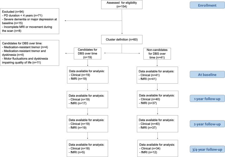
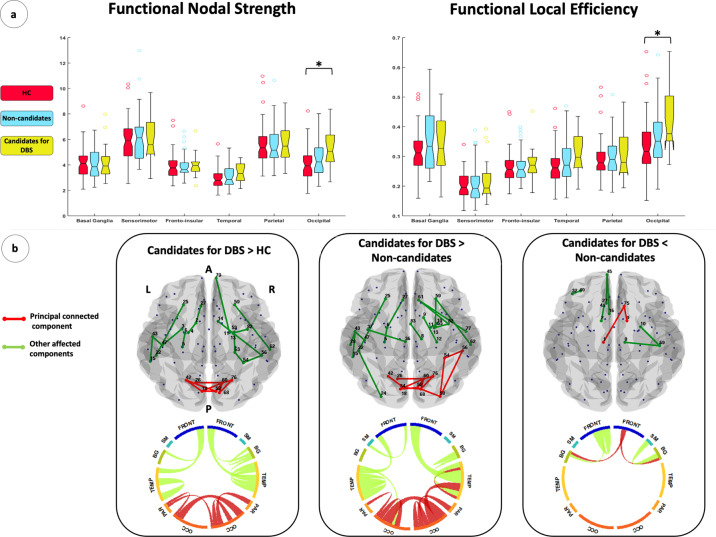
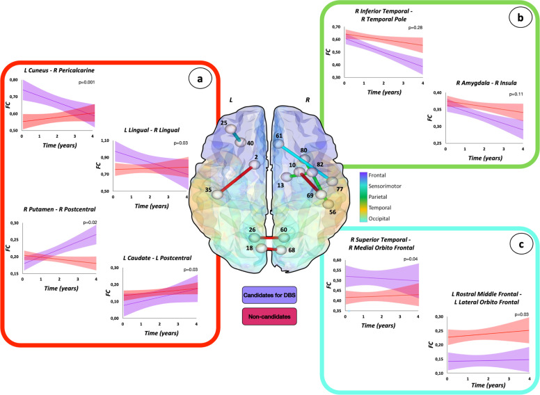
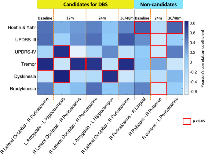
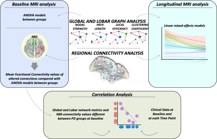
This study aimed to identify functional neuroimaging patterns anticipating the clinical indication for deep brain stimulation (DBS) in patients with Parkinson's disease (PD). A cohort of prospectively recruited patients with PD underwent neurological evaluations and resting-state functional MRI (RS-fMRI) at baseline and annually for 4 years. Patients were divided into two groups: 19 patients eligible for DBS over the follow-up and 41 patients who did not meet the criteria to undergo DBS. Patients selected as candidates for DBS did not undergo surgery at this stage. Sixty age- and sex-matched healthy controls performed baseline evaluations. Graph analysis and connectomics assessed global and local topological network properties and regional functional connectivity at baseline and at each time point. At baseline, network analysis showed a higher mean nodal strength, local efficiency, and clustering coefficient of the occipital areas in candidates for DBS over time relative to controls and patients not eligible for DBS. The occipital hyperconnectivity pattern was confirmed by regional analysis. At baseline, a decreased functional connectivity between basal ganglia and sensorimotor/frontal networks was found in candidates for DBS compared to patients not eligible for surgery. In the longitudinal analysis, patient candidate for DBS showed a progressively decreased topological brain organization and functional connectivity, mainly in the posterior brain networks, and a progressively increased connectivity of basal ganglia network compared to non-candidates for DBS. RS-fMRI may support the clinical indication to DBS and could be useful in predicting which patients would be eligible for DBS in the earlier stages of PD.


Conflict of interest statement

L.A., S.B., C.C., E.S., I.S., V.M., and P.M. declare no competing interests. F.A. is Section Editor of NeuroImage: Clinical; received compensation for consulting services and/or speaking activities from Philips, Biogen Idec, and Roche; and receives or has received research supports from the Italian Ministry of Health, AriSLA (Fondazione Italiana di Ricerca per la SLA), and the European Research Council. T.S and E.S. have received the Alzheimer Association grant for Promoting Diversity, from 2018 and honoraria from Actavis. A.T. has received honoraria for serving as a consultant to and on the advisory board of Univar BV. V.S.K. has received speaker honoraria from Roche and Alkaloid and receives research supports from the Swiss Pharm and Serbian Ministry of Education, Science, and Development and Serbian Academy of Sciences and Art. M.F. is Editor-in-Chief of the Journal of Neurology and Associate Editor of Human Brain Mapping; received compensation for consulting services and/or speaking activities from Almirall, Alexion, Bayer, Biogen, Celgene, Eli Lilly, Genzyme, Merck-Serono, Novartis, Roche, Sanofi, Takeda, and Teva Pharmaceutical Industries; and receives research support from Biogen Idec, Merck-Serono, Novartis, Roche, Teva Pharmaceutical Industries, Italian Ministry of Health, Fondazione Italiana Sclerosi Multipla, and ARiSLA (Fondazione Italiana di Ricerca per la SLA).

Figures

Fig. 1
Fig. 1. Flowchart illustrating the inclusion/exclusion of participants in the study.
Chart data for 154 patients with idiopathic Parkinson’s disease (PD) prospectively recruited at the Clinic of Neurology, Faculty of Medicine, University of Belgrade, Belgrade, Serbia. According to inclusion and exclusion criteria, 60 patients were included in the study. The study cohort was then divided in two groups: 19 patients eligible for DBS treatment and 41 patients who did not meet the criteria to undergo DBS over time. Patients were assessed by clinical, cognitive/behavioral, and brain MRI evaluations at study entry and every year for a maximum of 4 years. DBS deep brain stimulation, fMRI functional MRI, PD Parkinson’s disease.
Fig. 2
Fig. 2. Functional connectome alterations in Parkinson’s disease patients relative to healthy controls at baseline.
a Box plot of lobar functional nodal strength and local efficiency in healthy controls, patients eligible for DBS over time (Candidates for DBS), and patients not eligible for future DBS (Non-candidates). The black horizontal line in each box plot represents the median, the two lines just above and below the median represent the 25th and 75th percentiles, whiskers represent the minimum and maximum values, and all the dots outside the confidence interval are considered as outliers. *p < 0.05, Bonferroni-corrected for multiple comparisons. b Regional analysis results (NBS), from left to right: increased functional connectivity in candidates for DBS relative to controls; increased functional connectivity in Candidates for DBS relative to Non-candidates; decreased functional connectivity in Candidates for DBS relative to Non-candidates. In the upper part of the B section, abnormal functional connections are represented in a glass brain. In the lower part, lobar regions are arranged as a ring (the size of the regions are proportional to the number of the nodes included). The principal (or largest) connected components are shown in red; other connected components, not included in the principal connected components, are shown in green. Supplementary Table 2 reports affected functional connection values as well as corresponding statistical significance values. Supplementary Table 5 reports the names of each brain node with the corresponding number. A anterior, BG basal ganglia, Candidates for DBS patients eligible for DBS, FRONT frontal, HC healthy controls, L left, Non-candidates patients not candidate to DBS, OCC occipital, PAR parietal, P posterior, R right, SM sensorimotor, TEMP temporal.
Fig. 3
Fig. 3. Functional connectivity changes over time in patients eligible or not to DBS.
Three distinct patterns of progression are represented: a different trend of change between PD groups (connections in red); b similar trend of change (increase or decrease), with or without functional connectivity difference between the groups (connections in green); and c different but stable functional connectivity values over time in the two groups (connections in light blue). The effects of age, sex, levodopa equivalent daily dose at study entry, and changes of treatment over time were considered in the model. Figure reports selected findings. For further details see Supplementary Table 3. Supplementary Table 5 reports the names of each brain node with the corresponding number. Candidates for DBS patients eligible for DBS, FC functional connectivity, L left, Non-candidates patients not eligible for DBS, R right.
Fig. 4
Fig. 4. Clinical-MRI correlations between functional network properties and clinical data in each Parkinson’s disease group.
Each column shows functional brain proprieties and each row clinical data at baseline and at each time point. Color scale represents Pearson’s correlation coefficient. Red squares indicate statistical significance at a threshold of p < 0.05. For further details see Supplementary Table 4. Candidates for DBS patients eligible for DBS, L left, m months, Non-candidates patients not eligible for DBS, R right, UPDRS Unified Parkinson’s disease rating scale.
Fig. 5
Fig. 5. MRI processing pipeline.
Baseline MRI analysis. Global and lobar network topological metrics were compared between groups using ANOVA models. NBS analysis compared functional connectivity between groups. Mean functional connectivity values of the resulting altered connections were obtained and compared between groups using ANOVA models. Longitudinal MRI analysis. Linear mixed-effects models were implemented to investigate global and lobar network measures and functional connectivity changes over time. Correlation analysis. Partial correlations were assessed between baseline fMRI metrics (global and lobar network metrics and NBS-connectivity values, which were found to be different between PD groups) and clinical data at baseline and at each time point using Pearson’s correlation coefficient. NBS network-based statistics.

References

    1. Poewe W, et al. Parkinson disease. Nat. Rev. Dis. Prim. 2017;3:17013. - PubMed
    1. Damier P, Hirsch EC, Agid Y, Graybiel AM. The substantia nigra of the human brain. I. Nigrosomes and the nigral matrix, a compartmental organization based on calbindin D(28K) immunohistochemistry. Brain. 1999;122:1421–1436. - PubMed
    1. Schuepbach WM, et al. Neurostimulation for Parkinson’s disease with early motor complications. N. Engl. J. Med. 2013;368:610–622. - PubMed
    1. Follett KA, et al. Pallidal versus subthalamic deep-brain stimulation for Parkinson’s disease. N. Engl. J. Med. 2010;362:2077–2091. - PubMed
    1. Lhommee E, et al. Behavioural outcomes of subthalamic stimulation and medical therapy versus medical therapy alone for Parkinson’s disease with early motor complications (EARLYSTIM trial): secondary analysis of an open-label randomised trial. Lancet Neurol. 2018;17:223–231. - PubMed