. 2022 Jan 10;8(1):2.
doi: 10.1038/s41531-021-00270-y.

Thalamic white matter macrostructure and subnuclei volumes in Parkinson's disease depression

Affiliations

Thalamic white matter macrostructure and subnuclei volumes in Parkinson's disease depression

R Bhome et al. NPJ Parkinsons Dis. .

Abstract

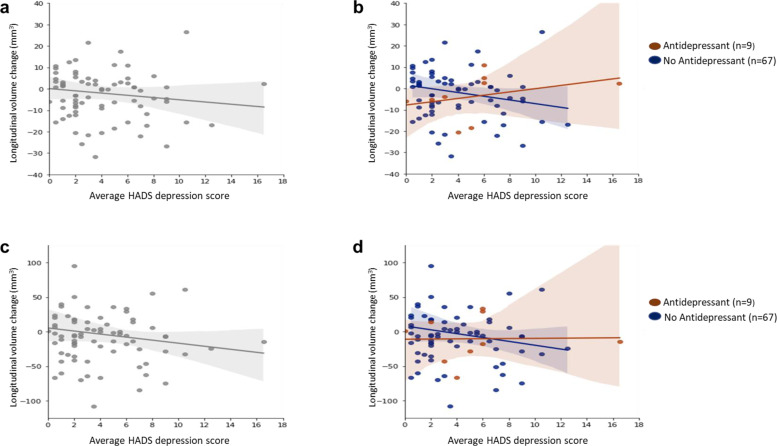
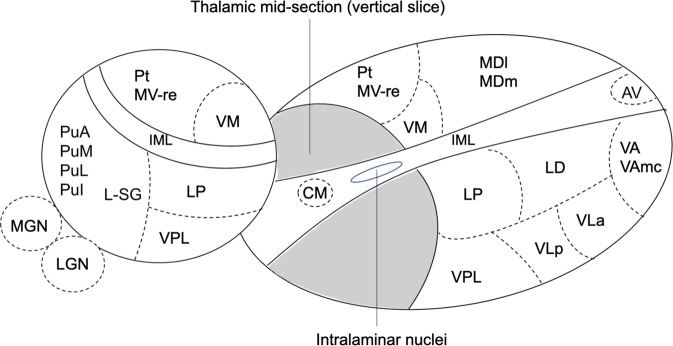
Depression is a common non-motor feature of Parkinson's disease (PD) which confers significant morbidity and is challenging to treat. The thalamus is a key component in the basal ganglia-thalamocortical network critical to the pathogenesis of PD and depression but the precise thalamic subnuclei involved in PD depression have not been identified. We performed structural and diffusion-weighted imaging (DWI) on 76 participants with PD to evaluate the relationship between PD depression and grey and white matter thalamic subnuclear changes. We used a thalamic segmentation method to divide the thalamus into its 50 constituent subnuclei (25 each hemisphere). Fixel-based analysis was used to calculate mean fibre cross-section (FC) for white matter tracts connected to each subnucleus. We assessed volume and FC at baseline and 14-20 months follow-up. A generalised linear mixed model was used to evaluate the relationship between depression, subnuclei volume and mean FC for each thalamic subnucleus. We found that depression scores in PD were associated with lower right pulvinar anterior (PuA) subnucleus volume. Antidepressant use was associated with higher right PuA volume suggesting a possible protective effect of treatment. After follow-up, depression scores were associated with reduced white matter tract macrostructure across almost all tracts connected to thalamic subnuclei. In conclusion, our work implicates the right PuA as a relevant neural structure in PD depression and future work should evaluate its potential as a therapeutic target for PD depression.


Conflict of interest statement

R.B., A.Z., G.E.C.T., J.E.I. and J.H.C. report no competing interests. R.S.W. has received speaker honoraria from GE Healthcare and honoraria from Britannia.

Figures

Fig. 1
Fig. 1. Relationship between HADS depression score and longitudinal change in pulvinar anterior and pulvinar medial volumes.
The top panels show scatter plots for longitudinal change in pulvinar anterior (PuA) volume in relation to average HADS depression score for all PD participants (a) and also separately for PD participants taking (brown) or not taking (blue) antidepressants (b). The corresponding scatter plots for pulvinar medial (PuM) volume change in relation to average HADS depression is shown in the bottom panels for all PD participants (c) and separately for participants taking (brown) or not taking (blue) antidepressants (d). Overall, higher HADS depression scores were associated with volume decreases in the right PuA (r = −0.12, P = 0.30) (a) and right PuM (r = −0.12, P = 0.28) (c). However, when participants were separated into those taking or not taking antidepressants, there was a positive correlation between HADS depression scores and longitudinal volume change in the right PuA for those taking antidepressants (r = 0.34, P = 0.37) but a negative correlation for those not taking antidepressants (r = −0.18, P = 0.14) (b). There was a similar pattern for the right PuM; Antidepressant group (r = 0.02, P = 0.96); No antidepressant group (r = −0.14, P = 0.26) (d).
Fig. 2
Fig. 2. Three-dimensional schematic representation of thalamic subnuclei.
The thalamus is divided into two by a vertical slice to reveal the medial surface. Abbreviations: Anterior nuclei (AV anteroventral); Lateral nuclei (LD laterodorsal, LP lateral posterior); Ventral nuclei (VA ventral anterior,VAmc ventral anterior magnocellular, VLa ventral lateral anterior, VLp ventral lateral posterior, VPL ventral posterolateral, VM ventromedial; Intralaminar nuclei (CM centromedian, CeM central medial, CL central lateral, Pc paracentral, Pf parafascicular);Medial nuclei (Pt paratenial, MV-re reuniens (medial ventral), MDm mediodorsal medial magnocellular, MDl mediodorsal lateral parvocellular); Posterior nuclei (LGN lateral geniculate nucleus, MGN medical geniculate nucleus, L-SG limitans (suprageniculate), PuA pulvinar anterior, PuM pulvinar medial, PuL pulvinar lateral, PuI pulvinar inferior); IML Internal medullary lamina.

References

    1. Reijnders JS, Ehrt U, Weber WE, Aarsland D, Leentjens AF. A systematic review of prevalence studies of depression in Parkinson’s disease. Mov. Disord. 2008;23:183–189. - PubMed
    1. Karlsen KH, Tandberg E, Arsland D, Larsen JP. Health related quality of life in Parkinson’s disease: a prospective longitudinal study. J. Neurol. Neurosurg. Psychiatry. 2000;69:584–589. - PMC - PubMed
    1. Jacob EL, Gatto NM, Thompson A, Bordelon Y, Ritz B. Occurrence of depression and anxiety prior to Parkinson’s disease. Parkinsonism Relat. Disord. 2010;16:576–581. - PMC - PubMed
    1. Srivastava A, et al. The relation between depression and parkin genotype: the CORE-PD study. Parkinsonism Relat. Disord. 2011;17:740–744. - PMC - PubMed
    1. Borgonovo J, et al. Changes in neural circuitry associated with depression at pre-clinical, pre-motor and early motor phases of Parkinson’s disease. Parkinsonism Relat. Disord. 2017;35:17–24. - PubMed