. 2022 Jan 10;12(1):468.
doi: 10.1038/s41598-021-04427-w.

Characterization of cisplatin-loaded chitosan nanoparticles and rituximab-linked surfaces as target-specific injectable nano-formulations for combating cancer

Affiliations

Characterization of cisplatin-loaded chitosan nanoparticles and rituximab-linked surfaces as target-specific injectable nano-formulations for combating cancer

Muhammad H Sultan et al. Sci Rep. .

Abstract

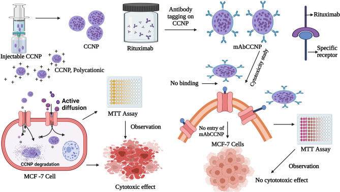
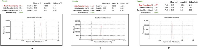
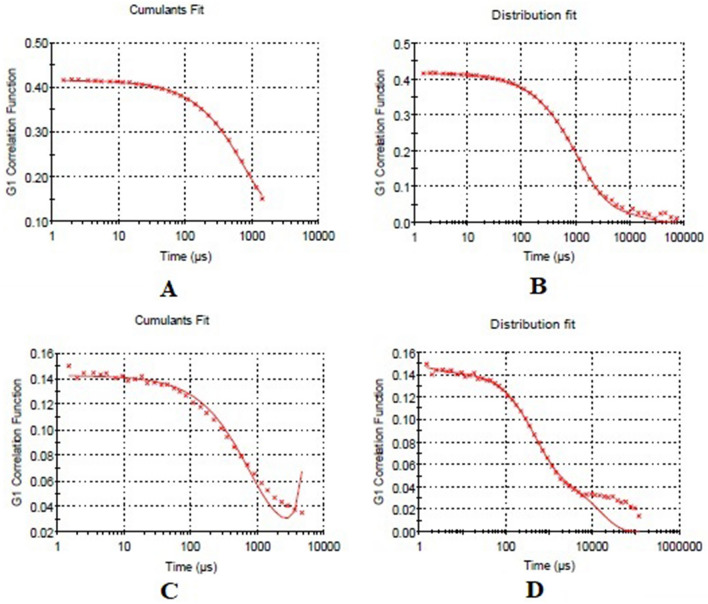
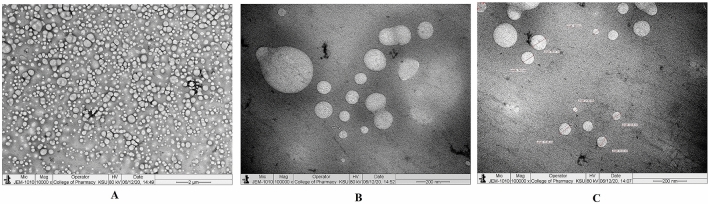
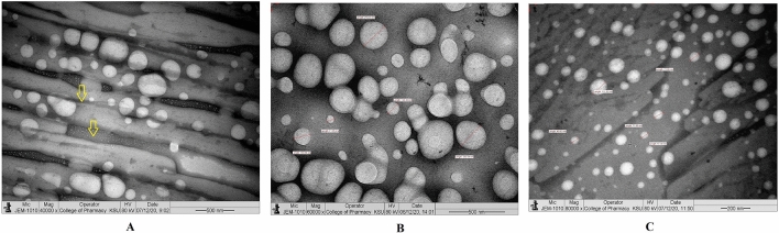
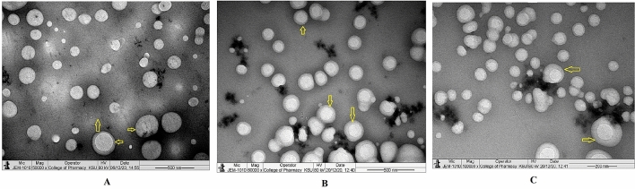
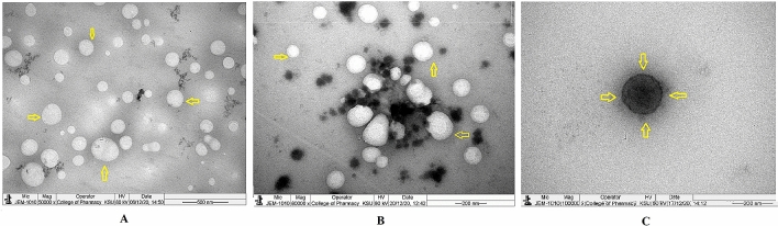
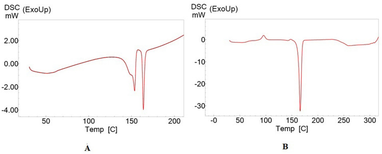
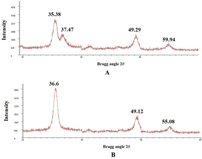
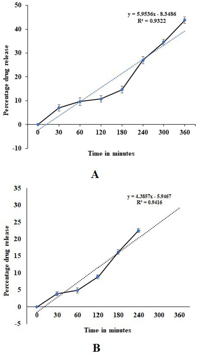
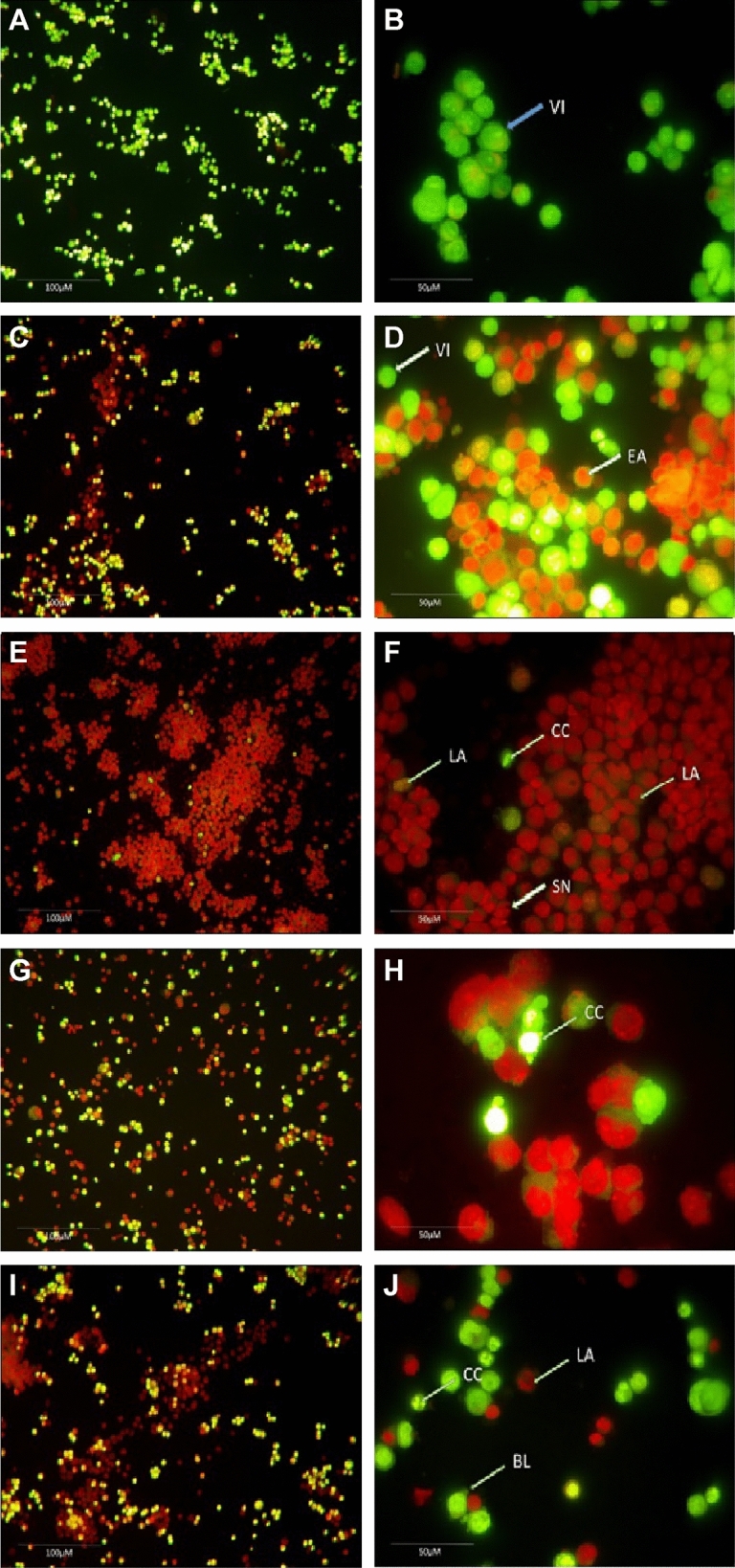
The present study was carried out to develop cisplatin-loaded chitosan nanoparticles (CCNP) and cisplatin-loaded chitosan nanoparticle surface linked to rituximab (mAbCCNP) as targeted delivery formulations. The two formulations (CCNP and mAbCCNP) exhibited significant physicochemical properties. The zetapotential (ZP) values of CCNP and mAbCCNP were 30.50 ± 5.64 and 26.90 ± 9.09 mV, respectively; while their particle sizes were 308.10 ± 1.10 and 349.40 ± 3.20 z.d.nm, respectively. The poly dispersity index (PDI) of CCNP was 0.257 ± 0.030 (66.6% PDI), while that of mAbCCNP was 0.444 ± 0.007 (57.60% PDI). Differential scanning calorimetry (DSC) revealed that CCNP had endothermic peaks at temperatures ranging from 135.50 to 157.69 °C. A sharp exothermic peak was observed at 95.79 °C, and an endothermic peak was observed at 166.60 °C. The XRD study on CCNP and mAbCCNP revealed distinct peaks at 2θ. Four peaks at 35.38°, 37.47°, 49.29°, and 59.94° corresponded to CCNP, while three distinct peaks at 36.6°, 49.12°, and 55.08° corresponded to mAbCCNP. The in vitro release of cisplatin from nanoparticles followed zero order kinetics in both CCNP and mAbCCNP. The profile for CCNP showed 43.80% release of cisplatin in 6 h (R2 = 0.9322), indicating linearity of release with minimal deviation. However, the release profile of mAbCCNP showed 22.52% release in 4 h (R2 = 0.9416), indicating linearity with sustained release. In vitro cytotoxicity studies on MCF-7 ATCC human breast cancer cell line showed that CCNP exerted good cytotoxicity, with IC50 of 4.085 ± 0.065 µg/mL. However, mAbCCNP did not elicit any cytotoxic effect. At a dose of 4.00 µg/mL cisplatin induced early apoptosis and late apoptosis, chromatin condensation, while it produced secondary necrosis at a dose of 8.00 µg/mL. Potential delivery system for cisplatin CCNP and mAbCCNP were successfully formulated. The results indicated that CCNP was a more successful formulation than mAbCCNP due to lack of specificity of rituximab against MCF-7 ATCC human breast cancer cells.


Conflict of interest statement

The authors declare no competing interests.

Figures

Figure 1
Figure 1
Schematic representation of nanoparticles developed and their mechanistic approach. *CCNP: Cisplatin loaded chitosan nanoparticles, **mAbCCNP: Rituximab surface linked cisplatin loaded chitosan nanoparticles. This figure was Created with BioRender.com, Bio Render, Canada.
Figure 2
Figure 2
Physical characterization of polymer, cross linker, and monoclonal antibody. (A) Zetapotential analysis of chitosan gel (1.0% w/v). (B) Zetapotential analysis of tripolyphosphate (0.5% w/v). (C) Zetapotential analysis of rituximab (1.0% v/v).
Figure 3
Figure 3
Physical characterization of nanoparticles. (A) Zetapotential analysis of injectable CCNP colloidal solution. (B) Size distribution analysis of injectable CCNP colloidal solution. (C) Zetapotential analysis of injectable mAbCCNP colloidal solution. (D) Size distribution analysis of injectable mAbCCNP colloidal solution.
Figure 4
Figure 4
Physical characterization of nanoparticles. (A) Cumulative fit analysis of injectable CCNP colloidal solution. (B) Size distribution analysis of injectable CCNP colloidal solution. (C) Cumulative fit analysis of injectable mAbCCNP colloidal solution. (D) Size distribution analysis of injectable mAbCCNP colloidal solution.
Figure 5
Figure 5
Transmission electron microscope study. (A) Morphology of injectable CCNP colloidal solution under ×10,000 magnification. (B) Morphology of injectable CCNP colloidal solution under ×100,000 magnification. (C) Morphology of injectable CCNP colloidal solution under ×100,000 magnification with size measurement.
Figure 6
Figure 6
Transmission electron microscope study. (A) Morphology of lyophilized CCNP under ×40,000 magnification. The yellow colour aero mark indicating fine lower size nanoparticles embedded in the mannitol powder which is representing the background. (B) Morphology of lyophilized CCNP under ×60,000 magnification with size measurement. (C) Morphology of lyophilized CCNP under ×80,000 magnification with size measurement.
Figure 7
Figure 7
Transmission electron microscope study. (A) Morphology of injectable mAbCCNP colloidal solution under ×50,000 magnification. (B) Morphology of injectable mAbCCNP colloidal solution under ×60,000 magnification (C) Morphology of injectable mAbCCNP colloidal solution under ×100,000 magnification. The yellow colour aero marks of all the three photographs indicating clear zone around the nanoparticle that represents rituximab attachment on CCNP.
Figure 8
Figure 8
Transmission electron microscope study. (A) Morphology of lyophilized mAbCCNP under ×50,000 magnification. (B) Morphology of lyophilized mAbCCNP under ×80,000 magnification. (C) Morphology of lyophilized mAbCCNP under ×100,000 magnification. The yellow colour aero marks of all the three photographs indicating clear zone around the nanoparticle that represents rituximab attachment on CCNP.
Figure 9
Figure 9
Differential Scanning Calorimetry analysis. (A) Thermogram of CCNP. (B) Thermogram of mAbCCNP.
Figure 10
Figure 10
X-ray diffractometer analysis. (A) Diffraction pattern of CCNP at . (B) Diffraction pattern of mAbCCNP at .
Figure 11
Figure 11
In vitro release profile study. (A) Release pattern of cisplatin from CCNP (B) Release pattern of cisplatin from mAbCCNP.
Figure 12
Figure 12
Morphological assessment of apoptotic development of MCF 7 cells by double staining technique using inverted phase contrast microscope. (A) Normal viable cells under ×20 magnification. (B) Normal viable cells under ×40 magnification. (C) Cisplatin treated at the concentration of 4 µg/mL under ×20 magnification. (D) Cisplatin treated at the concentration of 4 µg/mL under ×40 magnification, the arrow mark showing both viable cells and early development of apoptosis. (E) Cisplatin treated at the concentration of 8 µg/mL under ×20 magnification. (F) Cisplatin treated at the concentration of 8 µg/mL under ×40 magnification, the arrow mark showing both viable cells and late development of apoptosis, chromatin condensation and secondary necrosis development. (G) CCNP treated at the concentration of 3.5 µg/mL under 20 × magnification. (H) CCNP treated at the concentration of 3.5 µg/mL under 40 × magnification, the arrow mark showing the chromatin condensation. (I) CCNP treated at the concentration of 7 µg/mL under ×40 magnification. (J) CCNP treated at the concentration of 7 µg/mL under ×40 magnification, the arrow mark showing the late apoptosis, chromatin condensation and blebbing of the cell membrane. VI: Viable cells; EA: Early Apoptosis; LA: Late Apoptosis; CC: Chromatin condensation; SN: Secondary necrosis; BL: Blebbing of the cell membrane.

References

    1. Minko T, Rodriguez-Rodriguez L, Pozharov V. Nanotechnology approaches for personalized treatment of multidrug resistant cancers. Adv. Drug Deliv. Rev. 2013;65:1880–1895. doi: 10.1016/j.addr.2013.09.017. - DOI - PubMed
    1. Jing-yi W, Yu W, Xia M. Chitosan nanolayered cisplatin-loaded lipid nanoparticles for enhanced anticancer efficacy in cervical cancer. Nanoscale Res. Lett. 2016;11(524):1–8. doi: 10.1186/s11671-016-1698-9. - DOI - PMC - PubMed
    1. Raju, V., Chandrababu, R., Ramar, T. Chapter 12—Targeted Nanotherapeutics Based on Cancer Biomarkers, Multifunctional Systems for Combined Delivery, Biosensing and Diagnostics, 229–244 10.1016/B978-0-323-52725-5.00012-5 (Elsevier Publications, 2017).
    1. Safhi, M.M., et al. Chapter 8—Nanoparticle System for Anticancer Drug Delivery: Targeting to Overcome Multidrug Resistance, Multifunctional Systems for Combined Delivery, Biosensing and Diagnostics. 159–169 10.1016/B978-0-323-52725-5.00008-3 (Elsevier Publications, 2017).
    1. Zahreddine H, Borden KL. Mechanisms and insights into drug resistance in cancer. Front. Pharmacol. 2013;4:28. doi: 10.3389/fphar.2013.00028. - DOI - PMC - PubMed

Publication types