

Functional cortical localization of tongue movements using corticokinematic coherence with a deep learning-assisted motion capture system
- PMID: 35013521
- PMCID: PMC8748830
- DOI: 10.1038/s41598-021-04469-0
Functional cortical localization of tongue movements using corticokinematic coherence with a deep learning-assisted motion capture system
Abstract
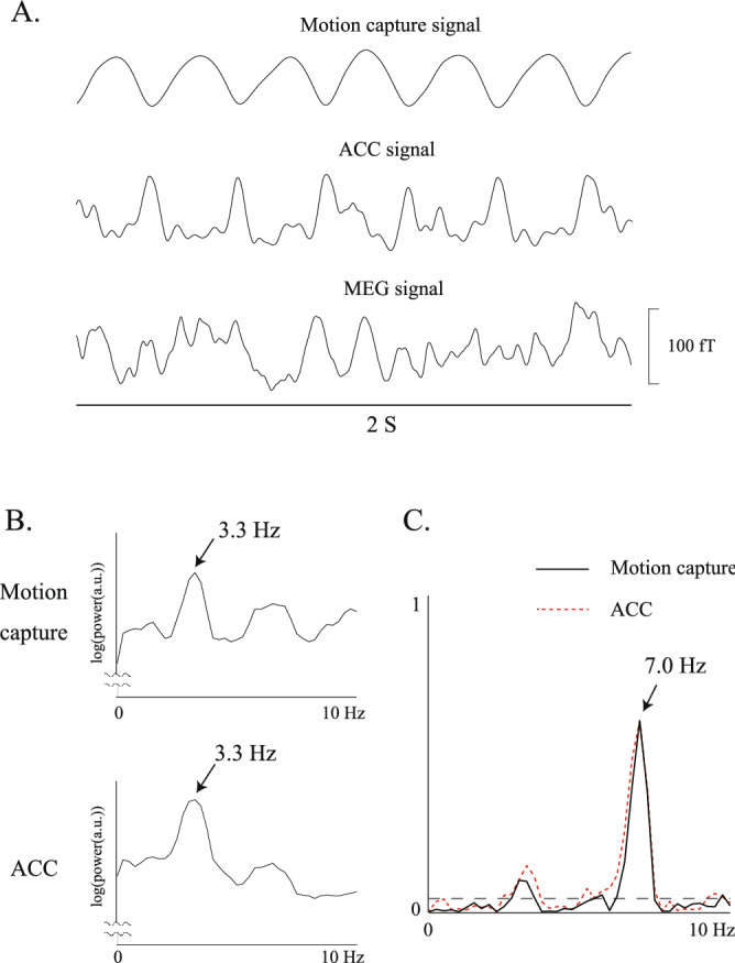
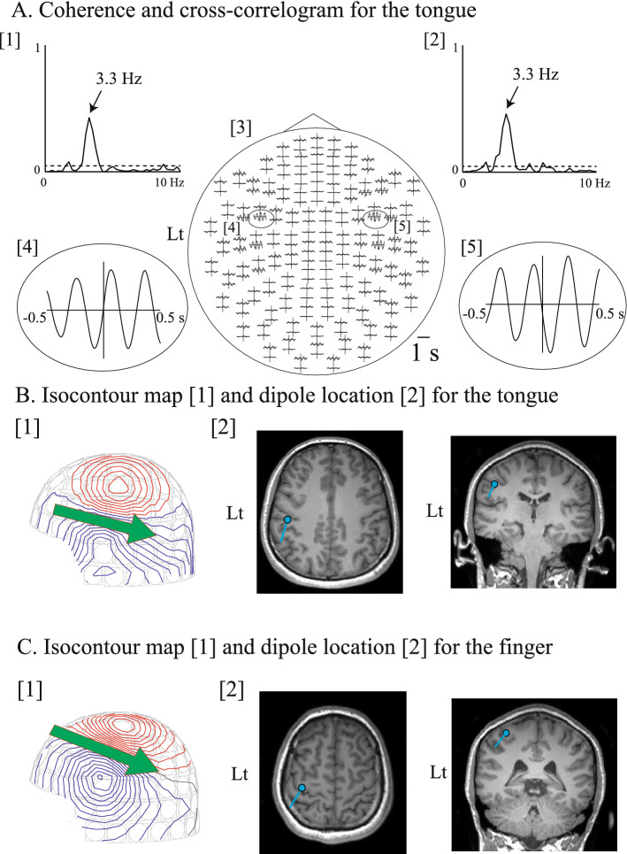
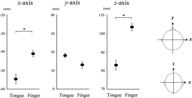
Corticokinematic coherence (CKC) between magnetoencephalographic and movement signals using an accelerometer is useful for the functional localization of the primary sensorimotor cortex (SM1). However, it is difficult to determine the tongue CKC because an accelerometer yields excessive magnetic artifacts. Here, we introduce a novel approach for measuring the tongue CKC using a deep learning-assisted motion capture system with videography, and compare it with an accelerometer in a control task measuring finger movement. Twelve healthy volunteers performed rhythmical side-to-side tongue movements in the whole-head magnetoencephalographic system, which were simultaneously recorded using a video camera and examined using a deep learning-assisted motion capture system. In the control task, right finger CKC measurements were simultaneously evaluated via motion capture and an accelerometer. The right finger CKC with motion capture was significant at the movement frequency peaks or its harmonics over the contralateral hemisphere; the motion-captured CKC was 84.9% similar to that with the accelerometer. The tongue CKC was significant at the movement frequency peaks or its harmonics over both hemispheres. The CKC sources of the tongue were considerably lateral and inferior to those of the finger. Thus, the CKC with deep learning-assisted motion capture can evaluate the functional localization of the tongue SM1.
© 2022. The Author(s).
Conflict of interest statement
The authors declare no competing interests.
Figures
Similar articles
-
More comprehensive proprioceptive stimulation of the hand amplifies its cortical processing.J Neurophysiol. 2022 Sep 1;128(3):568-581. doi: 10.1152/jn.00485.2021. Epub 2022 Jul 20. J Neurophysiol. 2022. PMID: 35858122 Free PMC article.
-
Proprioceptive response strength in the primary sensorimotor cortex is invariant to the range of finger movement.Neuroimage. 2023 Apr 1;269:119937. doi: 10.1016/j.neuroimage.2023.119937. Epub 2023 Feb 13. Neuroimage. 2023. PMID: 36791896
-
Corticokinematic coherence is stronger to regular than irregular proprioceptive stimulation of the hand.J Neurophysiol. 2021 Aug 1;126(2):550-560. doi: 10.1152/jn.00095.2021. Epub 2021 Jul 14. J Neurophysiol. 2021. PMID: 34259024
-
Coupling between human brain activity and body movements: Insights from non-invasive electromagnetic recordings.Neuroimage. 2019 Dec;203:116177. doi: 10.1016/j.neuroimage.2019.116177. Epub 2019 Sep 9. Neuroimage. 2019. PMID: 31513941 Review.
-
Sensorimotor Mapping With MEG: An Update on the Current State of Clinical Research and Practice With Considerations for Clinical Practice Guidelines.J Clin Neurophysiol. 2020 Nov;37(6):564-573. doi: 10.1097/WNP.0000000000000481. J Clin Neurophysiol. 2020. PMID: 33165229 Review.
Cited by
-
Neurophysiological Basis of Deep Brain Stimulation and Botulinum Neurotoxin Injection for Treating Oromandibular Dystonia.Toxins (Basel). 2022 Nov 2;14(11):751. doi: 10.3390/toxins14110751. Toxins (Basel). 2022. PMID: 36356002 Free PMC article. Review.
-
Cortico force coherence of the finger and toe with slight rhythmic pressure on force sensors using electroencephalography.Sci Rep. 2025 Apr 12;15(1):12639. doi: 10.1038/s41598-025-95759-4. Sci Rep. 2025. PMID: 40221443 Free PMC article.
-
A review of combined functional neuroimaging and motion capture for motor rehabilitation.J Neuroeng Rehabil. 2024 Jan 3;21(1):3. doi: 10.1186/s12984-023-01294-6. J Neuroeng Rehabil. 2024. PMID: 38172799 Free PMC article. Review.
References
-
- Penfield W, Boldrey E. Somatic motor and sensory representation in the cerebral cortex of man as studied by electrical stimulation. Brain. 1937;60:389–443. doi: 10.1093/brain/60.4.389. - DOI
Publication types
MeSH terms
Grants and funding
LinkOut - more resources
Full Text Sources
