. 2020 Mar 6:2020:6150942.
doi: 10.1155/2020/6150942. eCollection 2020.

Caveolin-1 Scaffolding Domain Peptide Regulates Colon Endothelial Cell Survival through JNK Pathway

Affiliations

Caveolin-1 Scaffolding Domain Peptide Regulates Colon Endothelial Cell Survival through JNK Pathway

Kai Fang et al. Int J Inflam. .

Abstract

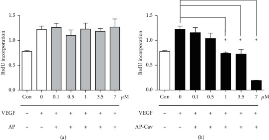
It has been reported that pathological angiogenesis contributes to both experimental colitis and inflammatory bowel disease. Recently, we demonstrated that endothelial caveolin-1 plays a key role in the pathological angiogenesis of dextran sodium sulfate (DSS) colitis. However, the molecular mechanism of caveolin-1 regulation of endothelial function is unknown. In this study, we examined how the antennapedia- (AP-) conjugated caveolin-1 scaffolding domain (AP-Cav) modulates vascular endothelial growth factor- (VEGF-) dependent colon endothelial cell angiogenic responses, as seen during colitis. We used mouse colon endothelial cells and found that AP-Cav significantly inhibited VEGF-mediated bromodeoxyuridine (BrdU) incorporation into colon microvascular endothelial cells. AP-Cav significantly blunted VEGF-dependent extracellular signal-regulated kinase 1/2 (ERK 1/2) phosphorylation at 10 minutes and 2 hours after stimulation, compared with the AP control peptide. AP-Cav + VEGF-A treatment also significantly increased c-Jun N-terminal kinase (JNK) phosphorylation at 2 hours. AP-Cav + VEGF-A treatment significantly downregulated retinoblastoma (Rb) protein levels, upregulated cleaved caspase-3 protein levels at 4 hours, and induced apoptosis. Thus, our study suggests that disruption of endothelial caveolin-1 function via the AP-Cav diverts VEGF signaling responses away from endothelial cell proliferation and toward apoptosis through the inhibition of mitogen-activated protein (MAP) kinase signaling and the induction of JNK-associated apoptosis.


Conflict of interest statement

The authors have no conflicts of interest.

Figures

Figure 1
Figure 1
AP-Cav inhibits mouse colon endothelial cell proliferation. (a) AP peptide pretreatment shows no effect on colon endothelial cell proliferation. (b) AP-Cav inhibits endothelial cell proliferation induced by VEGF. Cells were treated with peptide at indicated concentrations for 30 minutes before being treated with VEGF (50 ng/ml) for 4 hours. Data are mean ± s.e.m. from three independent experiments p < 0.05.
Figure 2
Figure 2
AP-Cav inhibits ERK1/2 phosphorylation. (a) After overnight low serum starvation, the sample was treated for 30 minutes with the AP-Cav peptide and then was stimulated with 50 ng VEGF/ml for 10 minutes. (b) After overnight low serum starvation, 30 minutes AP-Cav peptide treatment, and stimulation with 50 ng VEGF/ml for 2 hours, the ERK1/2 phosphorylation level was determined. Cells were treated with peptide for 30 minutes and then stimulated with 50 ng/ml of VEGF at the indicated times. At the end of the stimulation, cells were immediately lysed in RIPA buffer. The lysates were subjected to SDS-PAGE and western blot analysis with specific antibodies for phospho-ERK 1/2 and total ERK 1/2. Bar graph represents the phospho-ERK 1/2 to total ERK 1/2 ratio normalized to beta-actin levels. The data are mean ± s.e.m. from three independent experiments, p < 0.05.
Figure 3
Figure 3
Effect of AP-Cav on cell cycle protein level of p27kip1, Rb in mouse colon endothelial cells. (a) After overnight low serum starvation, 30 minute AP-Cav peptide treatment, and stimulation with 50 ng VEGF/ml for 0, 2, and 4 hours, cells were subjected to SDS-PAGE and western blot analysis with specific antibodies for p27kip1 or Rb. Beta-actin was used as a loading control. (b) Bar graph represents relative Rb level normalized to beta-actin level. (c) Bar graph represents relative level of p27kip1 normalized to beta-actin level. Data are mean ± s.e.m. from three independent experiments, p < 0.05.
Figure 4
Figure 4
AP-Cav peptide upregulates caspase-3 activity and JNK activity in mouse colon endothelial cells. (a) Mouse colon endothelial cells were starved in media containing 1% FBS overnight, treated with the peptide for 30 minutes, and stimulated with 50 ng/ml of VEGF for 0, 2, and 4 hours. At the end of the stimulation, cells were lysed in boiling SDS-sample buffer, and cell lysates were subjected to SDS-PAGE and western blot analysis with specific antibodies for the activated form of caspase-3 and phospho-JNK. The protein loading was probed with antibody for beta-actin. (b) Bar graph represents relative cleaved form of caspase-3 normalized to beta-actin level. (c) Bar graph represents phospho-JNK level normalized to beta-actin level. Data are mean ± s.e.m. from three independent experiments, p < 0.05.
Figure 5
Figure 5
JNK inhibition reverses the effect of AP-Cav on mouse colon endothelial cell apoptosis. Mouse colon endothelial cells were starved in media containing 1% FBS overnight, treated with JNK inhibitor for 1 hour, incubated for 30 minutes with AP or AP-Cav peptide, and stimulated with 50 ng/ml of VEGF for 4 hours. (a) At the end of the treatment, the cells were lysed in boiling SDS-sample buffer followed by western blot analysis with antibody for activated form of caspase-3. (b) Bar graph represents relative cleaved form of caspase-3 level normalized to beta-actin level. (c) At the end of the treatment, the cells were fixed and analyzed using fluorescein FragEL DNA fragmentation detection kit and then evaluated for apoptotic cells under a fluorescence microscope. The TUNEL positive cell number per field under 10x objective was counted. Data are mean ± s.e.m. from three independent experiments, p < 0.05.

Similar articles

References

    1. Chidlow J. H., Jr., Greer J. J. M., Anthoni C., et al. Endothelial caveolin-1 regulates pathologic angiogenesis in a mouse model of colitis. Gastroenterology . 2009;136(2):575–584.e2. doi: 10.1053/j.gastro.2008.10.085. - DOI - PMC - PubMed
    1. Danese S., Sans M., Spencer D. M., et al. Angiogenesis blockade as a new therapeutic approach to experimental colitis. Gut . 2007;56(6):855–862. doi: 10.1136/gut.2006.114314. - DOI - PMC - PubMed
    1. Filippini A., Sica G., D’Alessio A. The caveolar membrane system in endothelium: from cell signaling to vascular pathology. Journal of Cellular Biochemistry . 2018;119(7):5060–5071. doi: 10.1002/jcb.26793. - DOI - PubMed
    1. Labrecque L., Royal I., Surprenant D. S., Patterson C., Gingras D., Béliveau R. Regulation of vascular endothelial growth factor receptor-2 activity by caveolin-1 and plasma membrane cholesterol. Molecular Biology of the Cell . 2003;14(1):334–347. doi: 10.1091/mbc.e02-07-0379. - DOI - PMC - PubMed
    1. Okamoto T., Schlegel A., Scherer P. E., Lisanti M. P. Caveolins, a family of scaffolding proteins for organizing “preassembled signaling complexes” at the plasma membrane. Journal of Biological Chemistry . 1998;273(10):5419–5422. doi: 10.1074/jbc.273.10.5419. - DOI - PubMed