. 2022 Aug;100(8):1585-1601.
doi: 10.1002/jnr.25008. Epub 2022 Jan 11.

Pharmacological activation of the Sonic hedgehog pathway with a Smoothened small molecule agonist ameliorates the severity of alcohol-induced morphological and behavioral birth defects in a zebrafish model of fetal alcohol spectrum disorder

Affiliations

Pharmacological activation of the Sonic hedgehog pathway with a Smoothened small molecule agonist ameliorates the severity of alcohol-induced morphological and behavioral birth defects in a zebrafish model of fetal alcohol spectrum disorder

Derek F Burton et al. J Neurosci Res. 2022 Aug.

Abstract

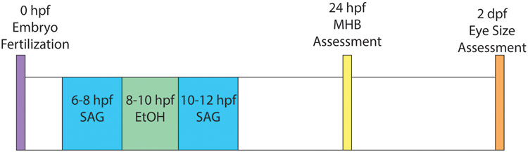
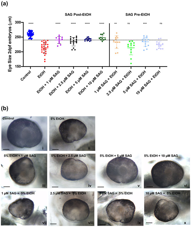
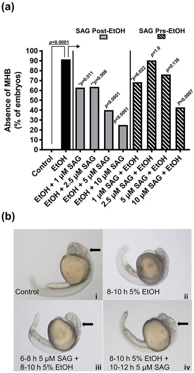
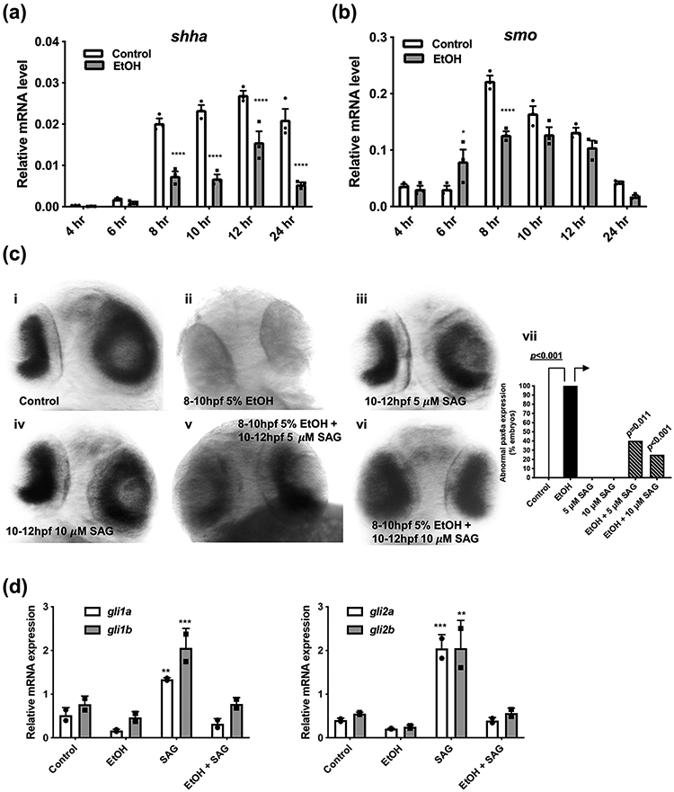
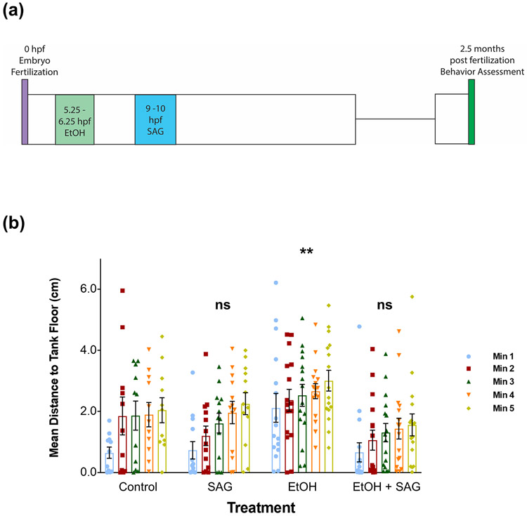
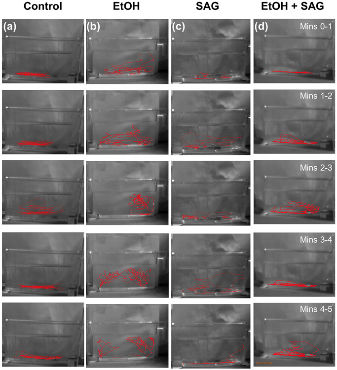
Ethanol exposure during the early stages of embryonic development can lead to a range of morphological and behavioral differences termed fetal alcohol spectrum disorders (FASDs). In a zebrafish model, we have shown that acute ethanol exposure at 8-10 hr postfertilization (hpf), a critical time of development, produces birth defects similar to those clinically characterized in FASD. Dysregulation of the Sonic hedgehog (Shh) pathway has been implicated as a molecular basis for many of the birth defects caused by prenatal alcohol exposure. We observed in zebrafish embryos that shh expression was significantly decreased by ethanol exposure at 8-10 hpf, while smo expression was much less affected. Treatment of zebrafish embryos with SAG or purmorphamine, small molecule Smoothened agonists that activate Shh signaling, ameliorated the severity of ethanol-induced developmental malformations including altered eye size and midline brain development. Furthermore, this rescue effect of Smo activation was dose dependent and occurred primarily when treatment was given after ethanol exposure. Markers of Shh signaling (gli1/2) and eye development (pax6a) were restored in embryos treated with SAG post-ethanol exposure. Since embryonic ethanol exposure has been shown to produce later-life neurobehavioral impairments, juvenile zebrafish were examined in the novel tank diving test. Our results further demonstrated that in zebrafish embryos exposed to ethanol, SAG treatment was able to mitigate long-term neurodevelopmental impairments related to anxiety and risk-taking behavior. Our results indicate that pharmacological activation of the Shh pathway at specific developmental timing markedly diminishes the severity of alcohol-induced birth defects.

Keywords: FASD; RRID:CVCL_0190; RRID:SCR_000441; RRID:SCR_002798; SAG; SMO; Shh; behavior; zebrafish.


Conflict of interest statement

CONFLICT OF INTEREST

The authors declare no competing financial interests.

Figures

Fig. 1.
Fig. 1.. Timeline schematic for ethanol and SAG treatment of zebrafish embryos.
Developmental timing (hpf, hours post fertilization; dpf, hours post fertilization) for ethanol exposure and SAG treatments are shown. Embryos are assessed for midbrain-hindbrain boundary (MHB) disruption at 24 hpf and eye size at 2 dpf.
Fig. 2.
Fig. 2.. SAG treatment after ethanol exposure rescues ocular size defects in 2 dpf zebrafish embryos.
(a) Zebrafish embryos were either pre-treated with SAG (1, 2.5, 5 or 10 μM) at 6-8 hpf and then exposed to 5% ethanol at 8-10 hpf (Pre-EtOH) or first exposed to 5% ethanol at 8-10 hpf and then treated with SAG (1, 2.5, 5 or 10 μM) at 10-12 hpf (Post-EtOH). Dotted line indicates threshold for normal eye size (> 240 μm). Eye diameter was measured at 2 dpf as described in Methods. n > 10 embryos per treatment. Error bars show mean ± SD. Data in (a) were evaluated by one-way ANOVA followed by Dunnett’s multiple comparisons test using GraphPad Prism version 8.02. Groups are compared to ethanol treated alone (EtOH). ANOVA summary was F(9,166) = 27.03, p<0.0001. Significant rescue is observed when SAG is given post-ethanol treatment (****p<0.0001 for all doses tested) with 90% of embryos having normal eye size at the 10 μM SAG treatment post-EtOH. In comparison, the rescue effect is less pronounced when SAG is given prior to EtOH, with 2 of the doses having no effect (**p=0.001 for 1 μM, ns (p>0.9999) for 2.5 μM, ***p=0.0002 for 5 μM and ns (p=0.2033) for 10 μM). See Supplemental Table 1 for summary of n and all multiple comparisons p values. (b) Representative images (i to x) for, controls, SAG + EtOH and EtOH + SAG exposures. The calibration bar indicates 50 μm.
Fig. 3.
Fig. 3.. SAG treatment mitigates the effects of acute ethanol exposure on MHB formation in zebrafish embryos.
Effects on MHB formation of treating embryos with SAG either Pre-EtOH (6-8 hpf) or Post-EtOH (10-12 hpf) exposure. (a) Zebrafish embryos ( n ≥ 10) were exposed to 5% ethanol at 8-10 hpf and treated with either 1, 2.5, 5 or 10 μM SAG pre- (SAG + EtOH) or post-EtOH (EtOH + SAG) exposure. Percent absence of MHB are shown; MHB absent in 0/35 control, 32/35 EtOH, 17/27 EtOH + 1 μM SAG, 21/33 EtOH + 2.5 μM SAG, 12/30 EtOH + 5 μM SAG, 3/12 EtOH + 10 μM SAG, 28/41 1 μM SAG + EtOH, 19/21 2.5 μM SAG + EtOH, 16/21 5 μM SAG + EtOH and 6/14 10 μM SAG + EtOH. Fisher’s exact test was used to compare absence of MHB between groups. We observed ~90% disruption of MHB with 5% EtOH exposure at 8-10 hpf (Fisher’s exact test, p<0.0001 compared to control), This effect was significantly reduced with all post-EtOH SAG treatments compared to EtOH alone (→), in particular with the 5 and 10 μM SAG treatments (Fisher’s exact test, both ****p<0.0001). SAG when given prior to ethanol (Pre-EtOH), was more variable and less effective on reducing ethanol-induced MHB loss, with the 2.5 and 5 μM treatments not significant, and the 1 and 10 μM SAG + EtOH treatments having a significant effect (Fisher’s exact test, *p=0.022 and ****p=0.0007 compared to EtOH alone, respectively) (Figure 3a). (b) Representative images for (i) control, (ii) EtOH (5%) alone, (iii) 6-8 hpf 5 μM SAG + EtOH and (iv) EtOH + 10-12 hpf 5 μM SAG. Arrow indicates defined MHB border. MHB was assessed at 24 hpf as described in Methods.
Fig. 4.
Fig. 4.. SAG treatment following ethanol exposure rescues Sonic hedgehog-mediated gene expression in 2 dpf zebrafish larvae.
Time course for shha (a) and smo (b) mRNA expression in zebrafish embryos (pooled embryos n≥10 per time point, 3 replicates/pooled group), either control or exposed to EtOH (5%) at 8-10 hpf. RNA was harvested at the indicated time points, quantified by qRT-PCR and normalized to an internal rpl13a mRNA control. Gene expression as described in Methods. Data were evaluated by two-way ANOVA followed by Sidak’s multiple comparisons test using GraphPad Prism version 8.02. For each time point, control compared to EtOH. (a) Shha: there was a significant interaction of treatment and time (F(5,24) = 15.65, p<0.0001). Ethanol exposure significantly reduced shh expression from 8 hr through to 24 hr by ~3-fold (****p <0.0001 for 8, 10, 12 and 24 hrs for control compared to EtOH, Figure 4a). (b) Smo: there was a significant interaction of treatment and time (F(5,24) = 11.43, p<0.0001). For ethanol exposure, only at the 6 hr (*p =0.0101) and 8 hr (****p<0.0001) time points is a decrease in smo seen. For the time period 10 to 24 hr there appeared to be no significant effect of ethanol exposure on smo expression. See Supplemental Table 1 for summary of all multiple comparisons p values. (c) Pax6 in situ mRNA expression in 2 dpf embryo retina. Representative images for (i) control embryos (15/15 (100% normal pax6a expression)). (ii) embryos exposed to 5% EtOH at 8-10 hpf (0/10,100% abnormal). Embryos treated at 10-12 hpf with (iii) 5 μM SAG alone (0/9, 0% abnormal) or (iv) 10 μM SAG alone (0/8, 0% abnormal). Embryos exposed to 5% EtOH at 8-10 hpf and then treated post-EtOH at 10-12 hpf with either (v) 5 μM SAG (6/10, 40% abnormal) or (vi) 10 μM SAG (3/12, 25% abnormal). (vii) Plot of % abnormal pax6a expression in embryo retina. Fisher’s exact test, p<0.001 EtOH compared to control. The incidence of abnormal pax6a expression in the embryo retina caused by EtOH was significantly altered with post-EtOH SAG treatments compared to EtOH alone (→), (Fisher’s exact test, *p=0.011 and ***p=0.0005 for 5 μM and 10 μM SAG, respectively). (d) Quantitation of gli1a/b and gli2a/b, mRNA expression by qRT-PCR in 2 dpf zebrafish embryos exposed to 5% EtOH and/or 10 μM SAG as indicated. Gene expression as described in Methods. n≥10 pooled embryos per treatment (2 replicates/pooled group). Data were evaluated by two-way ANOVA followed by Dunnett’s multiple comparisons test using GraphPad Prism version 8.02. Groups are compared to control. Gli1: ANOVA summary for treatment was F(3,8) = 38.0, p<0.0001. SAG treatment significantly increased gli1a (**p=0.0084) and gli1b (***p=0.0005) expression. SAG treatment post-EtOH reduces gli1a/b expression to levels comparable to control. Gli2: ANOVA summary for treatment was F(3,8) = 42.4, p<0.0001. SAG treatment significantly increased gli2a (***p=0.0006) and gli1b (**p=0.0010) expression. SAG treatment post-EtOH reduces gli2a/b expression to levels comparable to control. See Supplemental Table 1 for summary of all multiple comparisons p values.
Fig. 5.
Fig. 5.. Altered tank diving behavior in juvenile zebrafish previously exposed to acute ethanol as embryos is rescued with embryonic SAG treatment.
(a) Timing schematic for ethanol exposure (1% at 5.25-6.25 hpf) + SAG treatment (5 μM post-EtOH 9-10 hpf) and behavior assessment at 60 to 75 days post fertilization. (b) Juvenile zebrafish (60-75 days) were individually assessed for changes in novel tank diving response as determined by mean distance traveled from the tank floor over a 5 min time course. Novel tank diving test data were analyzed by two-way ANOVA statistical analysis using GraphPad Prism version 8.02. Total of n > 10 per group from two independent experiments. Each bar represents the mean distance from the tank floor for each 1 min (1-5 mins). Error bars represent SEM. To compare groups, the mean was taken across all 5 minutes for each exposure group. A significant main effect of treatment was observed (F (3, 260) = 13.79, p<0.0001). The Tukey post-hoc multiple comparisons test was used to determine differences between exposure groups (groups are compared to control). Altered tank diving response, an indication of risk-taking behavior, is only observed for juvenile fish previously exposed embryonically to 1% ethanol (significantly different from control (post hoc) across all time points analyzed, **p=0.0016). Embryonic SAG treatment alone did not significantly (ns) affect the novel tank diving behavior of the fish at the juvenile stage, p=0.9721). Treatment of embryos post-EtOH exposure with SAG (EtOH + SAG) is able to rescue the risk-taking behavior with values not significantly (ns) different compared to control, p=0.2241. See Supplemental Table 1 for summary of all n and multiple comparisons p values.
Fig. 6.
Fig. 6.. Novel tank diving tracking in juvenile zebrafish exposed to acute ethanol with and without SAG treatment as embryos.
Representative novel tank diving tracks of each minute from 5 min videos of juvenile zebrafish. From left to right; (a) control, (b) 1% EtOH alone at 5.25-6.25 hpf, (c) SAG treatment alone at 9-10 hpf, (d) 1% EtOH at 5.25-6.25 hpf followed by SAG treatment at 9-10 hpf. Swimming was tracked in real time by video and analyzed by EthoVision XT tracking software (Noldus, Netherlands) that calculates distance from the tank floor and total distance traveled. A minimum of 10 fish were assessed for each treatment group.

References

    1. Ahlgren SC, Thakur V, & Bronner-Fraser M (2002). Sonic hedgehog rescues cranial neural crest from cell death induced by ethanol exposure. Proceedings of the National Academy of Sciences, 99(16), 10476–10481. - PMC - PubMed
    1. Albert S, Müller F, Fischer N, Biellmann D, Neumann C, Blader P, & Strähle U (2003). Cyclops-independent floor plate differentiation in zebrafish embryos. Developmental dynamics: an official publication of the American Association of Anatomists, 226(1), 59–66. - PubMed
    1. Aoto K, Shikata Y, Higashiyama D, Shiota K, & Motoyama J (2008). Fetal ethanol exposure activates protein kinase A and impairs Shh expression in prechordal mesendoderm cells in the pathogenesis of holoprosencephaly. Birth Defects Research Part A: Clinical and Molecular Teratology, 82(4), 224–231. - PubMed
    1. Arenzana F, Carvan MJ III, Aijon J, Sanchez-Gonzalez R, Arevalo R, & Porteros A (2006). Teratogenic effects of ethanol exposure on zebrafish visual system development. Neurotoxicology and teratology, 28(3), 342–348. - PubMed
    1. Arsić D, Beasley SW, & Sullivan MJ (2007). Switched-on Sonic hedgehog: A gene whose activity extends beyond fetal development–to oncogenesis. Journal of paediatrics and child health, 43(6), 421–423. - PubMed

Publication types