. 2022 Feb;25(2):72.
doi: 10.3892/mmr.2022.12588. Epub 2022 Jan 11.

Inhibition of FOXO1‑mediated autophagy promotes paclitaxel‑induced apoptosis of MDA‑MB‑231 cells

Affiliations

Inhibition of FOXO1‑mediated autophagy promotes paclitaxel‑induced apoptosis of MDA‑MB‑231 cells

Kaixiang Xu et al. Mol Med Rep. 2022 Feb.

Abstract

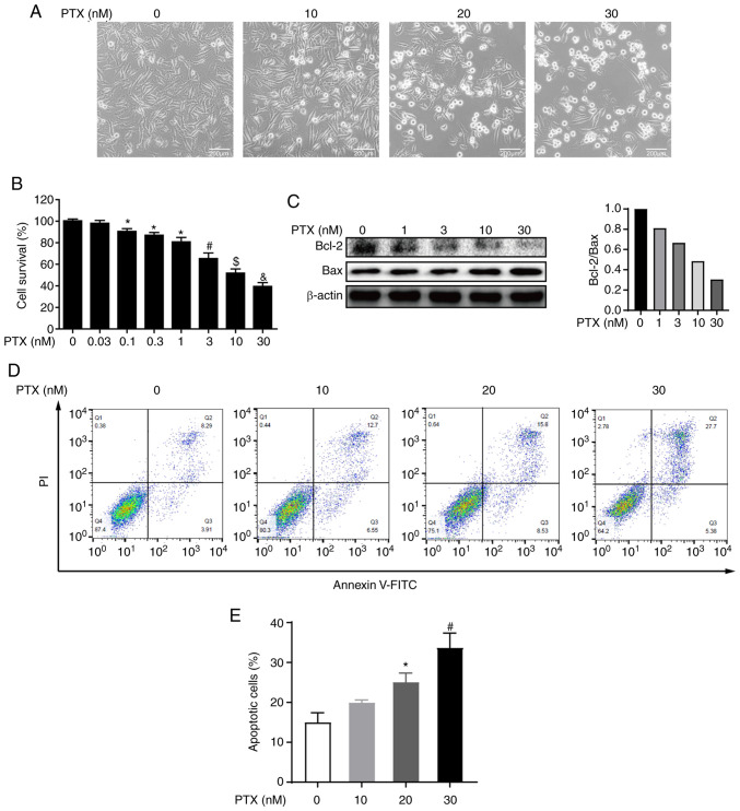
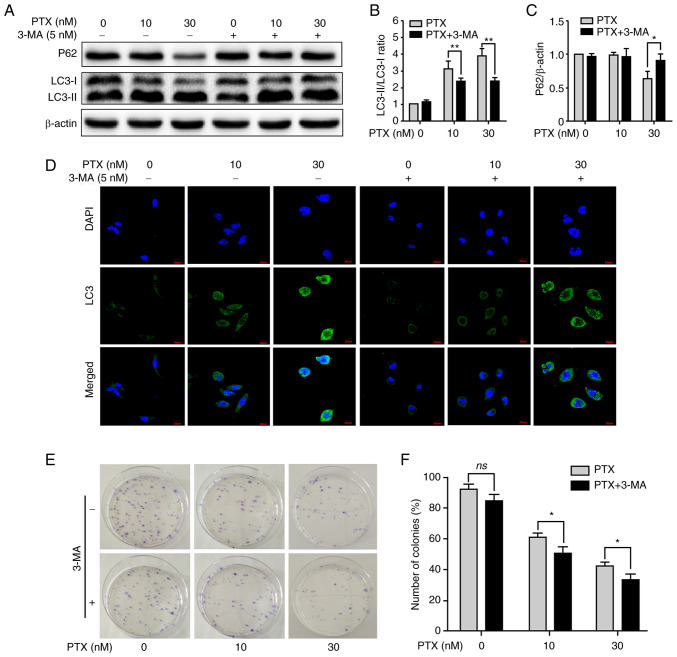
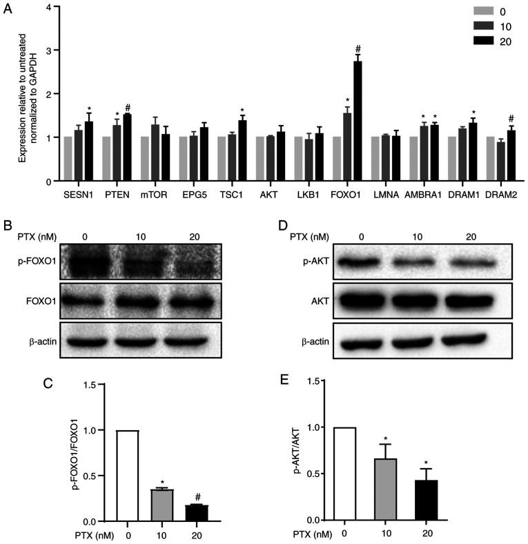
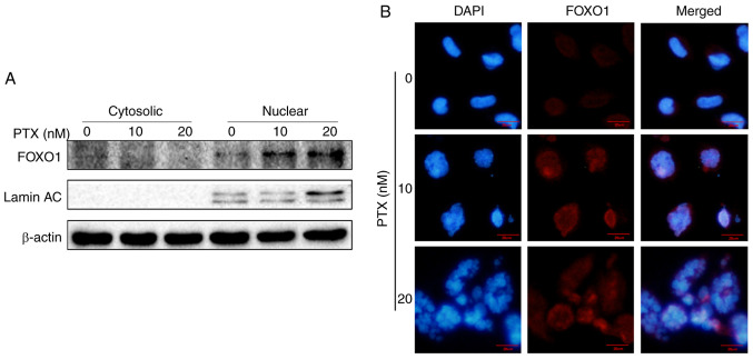
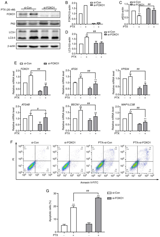
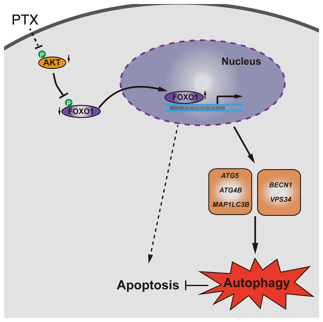
Triple‑negative breast cancer (TNBC) is the most aggressive subtype of breast cancer, and it often becomes resistant to paclitaxel (PTX) therapy. Autophagy plays an important cytoprotective role in PTX‑induced tumor cell death, and targeting autophagy has been promising for improving the efficacy of tumor chemotherapy in recent years. The aim of the present study was to clarify the mechanism of PTX inducing autophagy in TNBC cells to provide a potential clinical chemotherapy strategy of PTX for TNBC. The present study reported that PTX induced both apoptosis and autophagy in MDA‑MB‑231 cells and that inhibition of autophagy promoted apoptotic cell death. Furthermore, it was found that forkhead box transcription factor O1 (FOXO1) enhanced PTX‑induced autophagy through a transcriptional activation pattern in MDA‑MB‑231 cells, which was associated with the downstream target genes autophagy related 5, class III phosphoinositide 3‑kinase vacuolar protein sorting 34, autophagy related 4B cysteine peptidase, beclin 1 and microtubule associated protein 1 light chain 3β. Knocking down FOXO1 attenuated the survival of MDA‑MB‑231 cells in response to PTX treatment. These findings may be beneficial for improving the treatment efficacy of PTX and to develop autophagic targeted therapy for TNBC.

Keywords: apoptosis; autophagy; forkhead box transcription factor O1; paclitaxel; triple‑negative breast cancer.


Conflict of interest statement

The authors declare that they have no competing interests.

Figures

Figure 1.
Figure 1.
PTX exerts cytotoxicity and induces apoptosis in MDA-MB-231 cells. (A) Images of MDA-MB-231 cells exposed to 0, 10, 20 and 30 nM PTX for 24 h (scale bar, 200 µm). (B) Quantification of cell viability after treatment with different doses of PTX. *P<0.05 vs. 0 nM; #P<0.05 vs. 1 nM PTX; $P<0.05 vs. 3 nM PTX; &P<0.05 vs. 10 nM PTX. (C) Effect of different doses of PTX on expression levels of the apoptosis proteins Bcl-2 and Bax, with β-actin as an internal control (left) and semi-quantification of the Bcl-2/Bax ratio (right). (D) Effect of different doses of PTX on the apoptosis of MDA-MB-231 cells. (E) Quantification of the ratio of apoptotic cells in response to different doses of PTX. *P<0.05 vs. 0 nM PTX; #P<0.05 vs. 20 nM PTX. PTX, paclitaxel.
Figure 2.
Figure 2.
PTX induces autophagy in MDA-MB-231 cells. (A) Protein expression levels of the autophagy markers P62, LC3-I and LC3-II at different doses of PTX. β-actin was used as an internal control. The semi-quantification of (B) P62 (*P<0.05 vs. 0 nM PTX), and (C) LC3-II/LC3-I protein expression (*P<0.05 vs. 0 nM PTX). (D) Representative images of LC3 puncta. Cells were stained with antibodies against LC3 (green), and nuclei were stained with DAPI (blue). Scale bar, 20 µm. (E) Quantification of LC3 puncta (**P<0.01 vs. 0 nM PTX; ##P<0.01 vs. 20 nM PTX). PTX, paclitaxel; LC3, light chain 3; Baf A1, bafilomycin A1.
Figure 3.
Figure 3.
Inhibition of autophagy with 3-MA promotes PTX-induced apoptotic cell death. (A) Effect of 3-MA on protein expression levels of the autophagy markers P62, LC3-I and LC3-II. β-actin was used as an internal control. Semi-quantification of the (B) LC3-II/LC3-I ratio and (C) P62 protein expression. (D) Representative images of LC3 puncta after treatment with or without 3-MA and/or PTX. (E) Detection of cell viability by colony formation assay. (F) Quantification of the cellular colony formation rate shown in E. Data are presented as the means ± SD of three independent experiments. *P<0.05 and **P<0.01; ns, non-significant; PTX, paclitaxel; LC3, light chain 3; 3-MA, 3-methyladenine.
Figure 4.
Figure 4.
Autophagy regulatory pathway induction by PTX in MDA-MB-231 cells. (A) mRNA expression levels of autophagy signaling pathway-related genes in response to treatment with 10 and 20 nM PTX. GAPDH was used as an internal control. (*P<0.05 vs. 0 nM PTX; #P<0.05 vs. 10 nM PTX). (B) Protein expression of FOXO1 and p-FOXO1 and (C) semi-quantification of the p-FOXO1/FOXO1 ratio (*P<0.05 vs. 0 nM PTX; #P<0.05 vs. 10 nM PTX). (D) Protein expression of AKT and p-AKT and (E) semi-quantification of the p-AKT/AKT ratio (*P<0.05 vs. 0 nM PTX). β-actin was used as an internal control. Data are presented as the means ± SD of three independent experiments. PTX, paclitaxel; FOXO1, forkhead box transcription factor O1; p-, phosphorylated; SESN1, sestrin 1; PTEN, phosphatase and tensin homolog; mTOR, mechanistic target of rapamycin kinase; EPG5, ectopic P-granules autophagy protein 5 homolog; TSC1, TSC complex subunit 1; AKT, serine/threonine kinase 1; LKB1, serine/threonine kinase 11; LMNA, lamin A/C; AMBRA1, autophagy and beclin 1 regulator 1; DRAM1, DNA damage regulated autophagy modulator 1; DRAM2, DNA damage regulated autophagy modulator 2.
Figure 5.
Figure 5.
PTX induces increased nuclear FOXO1 in MDA-MB-231 cells. (A) Localization of FOXO1 by western blotting. Lamin AC, a nucleus-specific marker, and β-actin were used as internal controls. (B) Localization of FOXO1 was detected by immunofluorescence staining (scale bar, 20 µm). PTX, paclitaxel; FOXO1, forkhead box transcription factor O1.
Figure 6.
Figure 6.
Knockdown of FOXO1 attenuates PTX-induced autophagy and promotes apoptotic cell death in MDA-MB-231 cells. (A) Protein expression of the autophagy markers P62, LC3-I and LC3-II after treatment with si-FOXO1, with β-actin as an internal control. Semi-quantification of (B) FOXO1, (C) P62 and (D) LC3-II protein expression. (E) The mRNA expression levels of FOXO1 and its downstream target genes after treatment with siRNA and/or PTX. (F) The effect of FOXO1 knockdown on PTX-induced apoptosis was analyzed by flow cytometry. (G) Quantification of the ratio of apoptotic cells after the knockdown of FOXO1. PTX was used at a concentration of 20 nM. Data are presented as the means ± SD of three independent experiments. *P<0.05 vs. 0 nM PTX; **P<0.01 vs. 0 nM PTX; #P<0.05 vs. 20 nM PTX + si-Con; ##P<0.01 vs. 20 nM PTX + si-Con. PTX, paclitaxel; FOXO1, forkhead box transcription factor O1; LC3, light chain 3; si, small interfering; ATG5, autophagy related 5; VPS34, class III phosphoinositide 3-kinase vacuolar protein sorting 34; ATG4B, autophagy related 4B cysteine peptidase; BECN1, beclin 1; MAP1LC3B, microtubule associated protein 1 light chain 3β.
Figure 7.
Figure 7.
Schematic model of FOXO1-mediated autophagy and apoptosis induced by PTX in MDA-MB-231 cells. PTX, paclitaxel; FOXO1, forkhead box transcription factor O1; p-, phosphorylated; ATG5, autophagy related 5; MAP1LC3B, microtubule associated protein 1 light chain 3β; BECN1, beclin 1; VPS34, class III phosphoinositide 3-kinase vacuolar protein sorting 34; ATG4B, autophagy related 4B cysteine peptidase.

Similar articles

Cited by

References

    1. Bianchini G, Balko JM, Mayer IA, Sanders ME, Gianni L. Triple-negative breast cancer: Challenges and opportunities of a heterogeneous disease. Nat Rev Clin Oncol. 2016;13:674–690. doi: 10.1038/nrclinonc.2016.66. - DOI - PMC - PubMed
    1. Mustacchi G, De Laurentiis M. The role of taxanes in triple-negative breast cancer: Literature review. Drug Des Devel Ther. 2015;9:4303–4318. doi: 10.2147/DDDT.S86105. - DOI - PMC - PubMed
    1. Yang N, Wang C, Wang J, Wang Z, Huang D, Yan M, Kamran M, Liu Q, Xu B. Aurora kinase A stabilizes FOXM1 to enhance paclitaxel resistance in triple-negative breast cancer. J Cell Mol Med. 2019;23:6442–6453. doi: 10.1111/jcmm.14538. - DOI - PMC - PubMed
    1. Lock R, Kenific CM, Leidal AM, Salas E, Debnath J. Autophagy-dependent production of secreted factors facilitates oncogenic RAS-driven invasion. Cancer Discov. 2014;4:466–479. doi: 10.1158/2159-8290.CD-13-0841. - DOI - PMC - PubMed
    1. Wu CC, Chan ML, Chen WY, Tsai CY, Chang FR, Wu YC. Pristimerin induces caspase-dependent apoptosis in MDA-MB-231 cells via direct effects on mitochondria. Mol Cancer Ther. 2005;4:1277–1285. doi: 10.1158/1535-7163.MCT-05-0027. - DOI - PubMed

MeSH terms