

Paracrine Wnt signaling is necessary for prostate epithelial proliferation
- PMID: 35014711
- PMCID: PMC8866211
- DOI: 10.1002/pros.24298
Paracrine Wnt signaling is necessary for prostate epithelial proliferation
Abstract
Introduction: The Wnt proteins play key roles in the development, homeostasis, and disease progression of many organs including the prostate. However, the spatiotemporal expression patterns of Wnt proteins in prostate cell lineages at different developmental stages and in prostate cancer remain inadequately characterized.
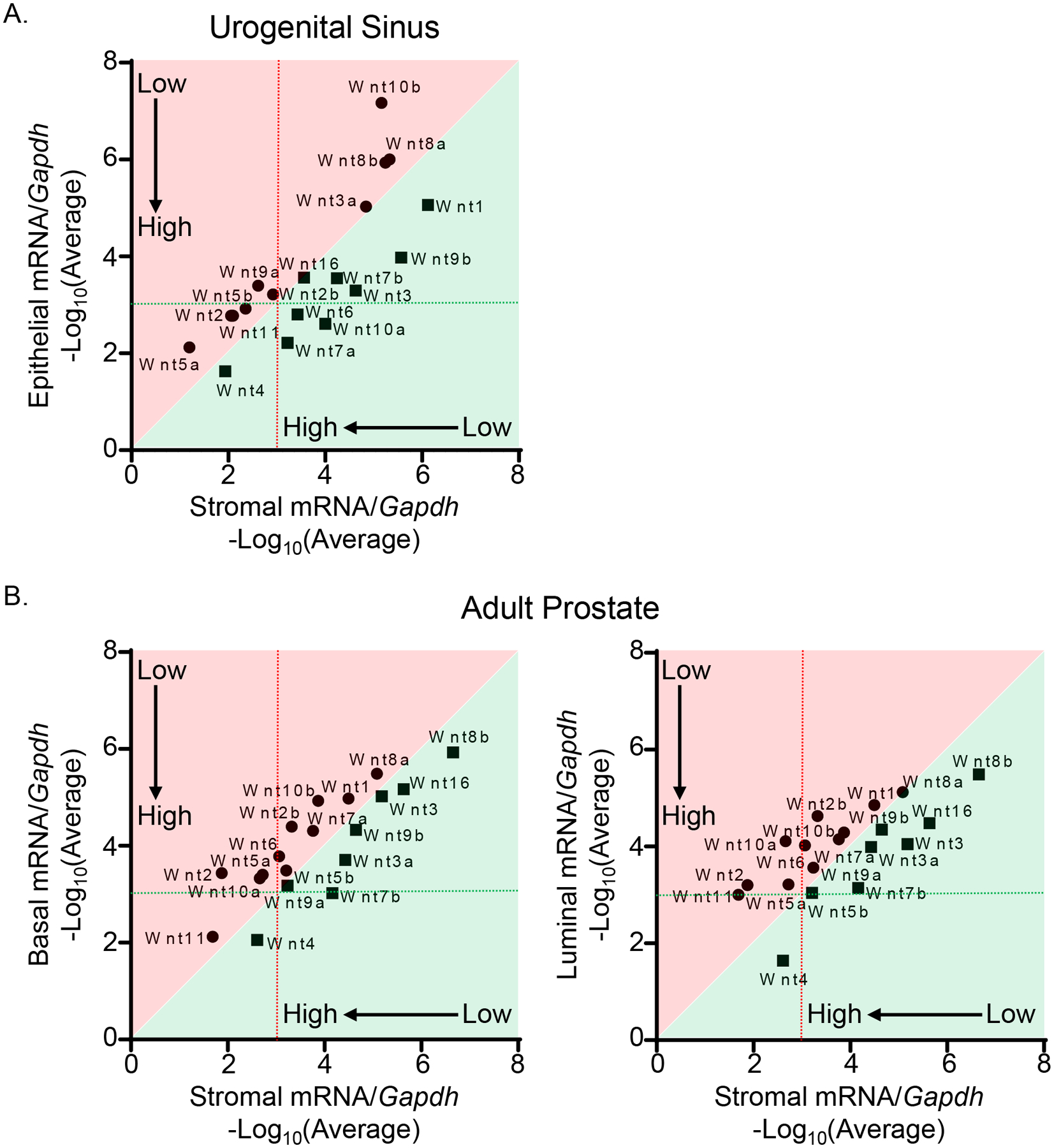
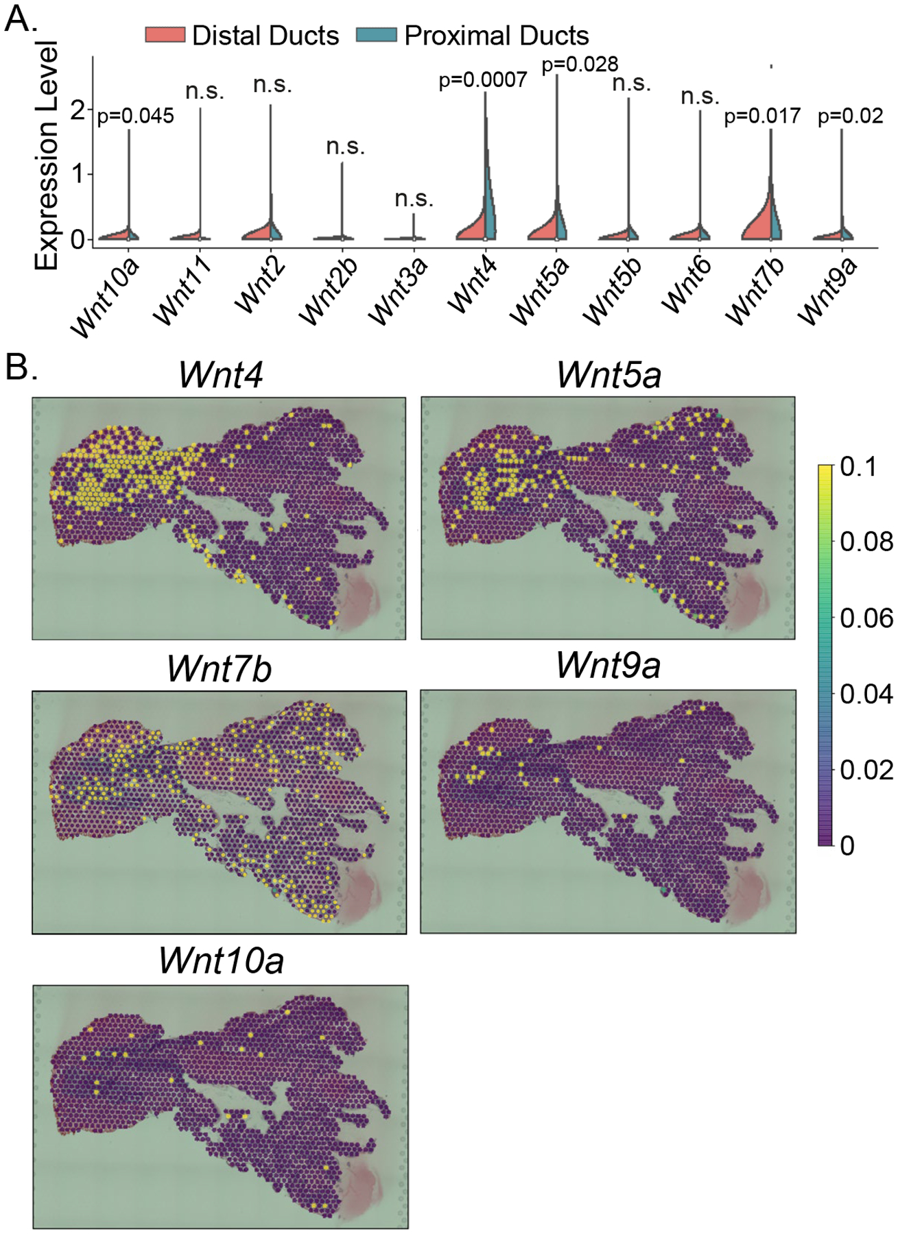
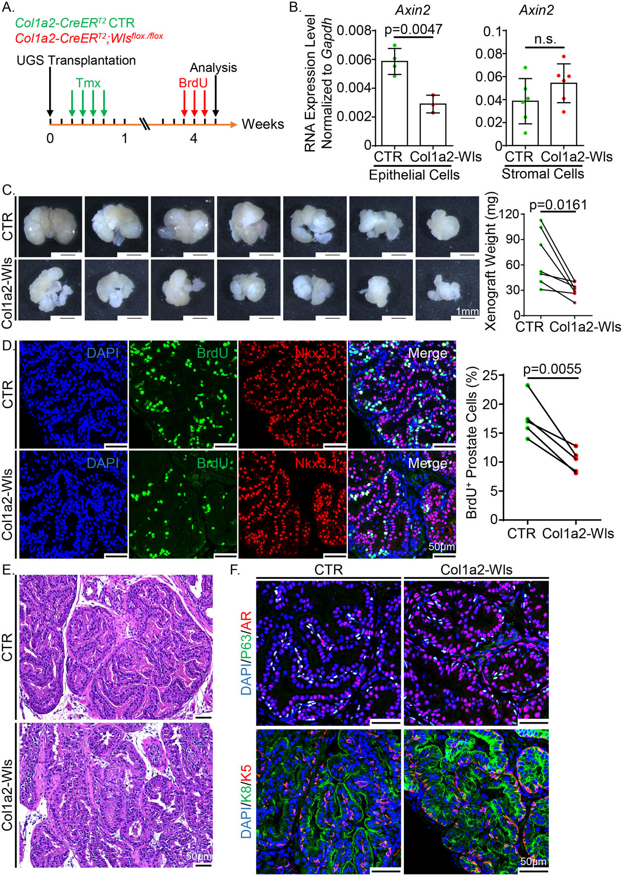
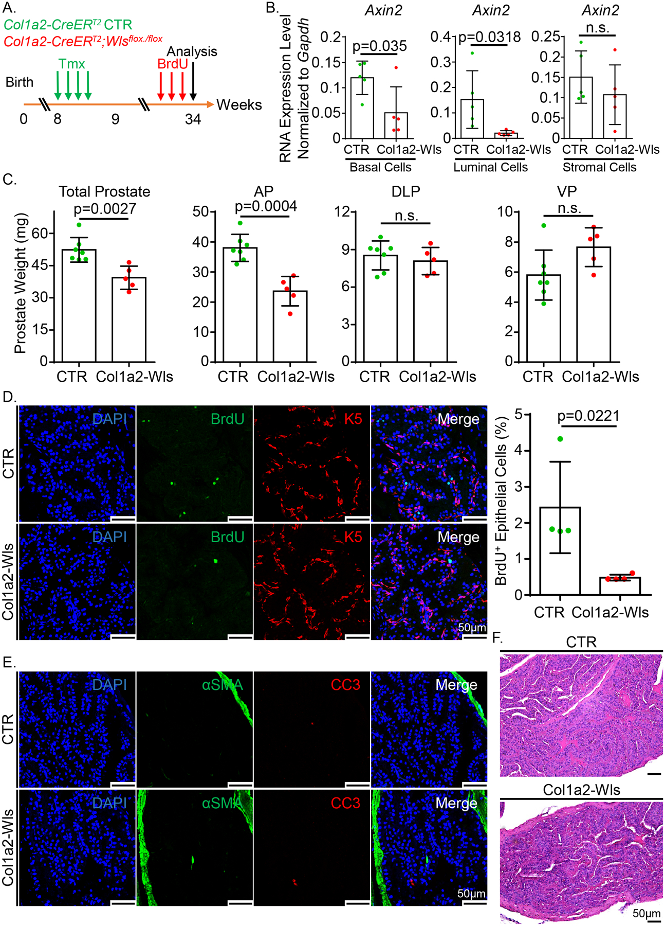
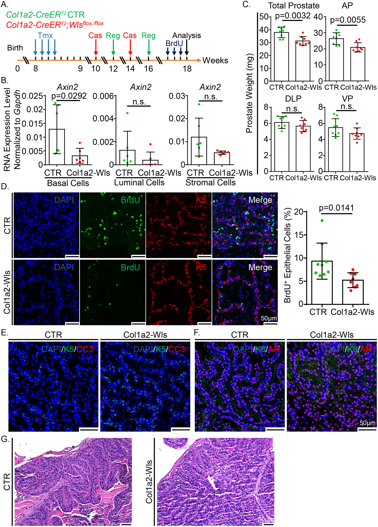
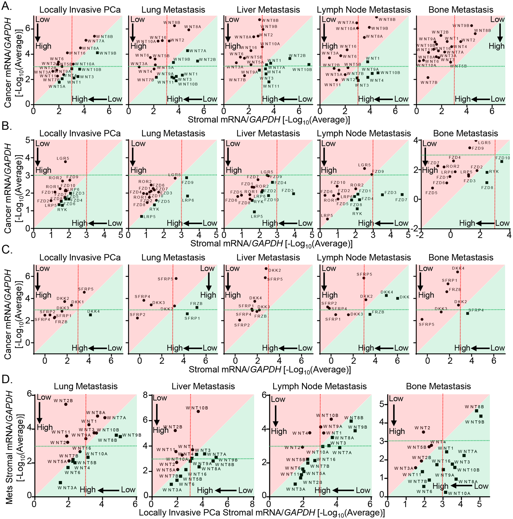
Methods: We isolated the epithelial and stromal cells in the developing and mature mouse prostate by flow cytometry and determined the expression levels of Wnt ligands. We used Visium spatial gene expression analysis to determine the spatial distribution of Wnt ligands in the mouse prostatic glands. Using laser-capture microscopy in combination with gene expression analysis, we also determined the expression patterns of Wnt signaling components in stromal and cancer cells in advanced human prostate cancer specimens. To investigate how the stroma-derived Wnt ligands affect prostate development and homeostasis, we used a Col1a2-CreERT2 mouse model to disrupt the Wnt transporter Wntless specifically in prostate stromal cells.
Results: We showed that the prostate stromal cells are a major source of several Wnt ligands. Visium spatial gene expression analysis revealed a distinct spatial distribution of Wnt ligands in the prostatic glands. We also showed that Wnt signaling components are highly expressed in the stromal compartment of primary and advanced human prostate cancer. Blocking stromal Wnt secretion attenuated prostate epithelial proliferation and regeneration but did not affect cell survival and lineage maintenance.
Discussion: Our study demonstrates a critical role of stroma-derived Wnt ligands in prostate development and homeostasis.
Keywords: Wls; Wnt; homeostasis; prostate cancer; prostate stromal cells.
© 2022 Wiley Periodicals LLC.
Conflict of interest statement
Conflicts of Interest
The authors declare no conflict of interest.
Figures
Similar articles
-
Identification of SFRP1 as a candidate mediator of stromal-to-epithelial signaling in prostate cancer.Cancer Res. 2005 Nov 15;65(22):10423-30. doi: 10.1158/0008-5472.CAN-05-0824. Cancer Res. 2005. PMID: 16288033
-
Dihydrotestosterone inhibits arylsulfatase B and Dickkopf Wnt signaling pathway inhibitor (DKK)-3 leading to enhanced Wnt signaling in prostate epithelium in response to stromal Wnt3A.Prostate. 2019 May;79(7):689-700. doi: 10.1002/pros.23776. Epub 2019 Feb 22. Prostate. 2019. PMID: 30801800
-
Stromal epigenetic dysregulation is sufficient to initiate mouse prostate cancer via paracrine Wnt signaling.Proc Natl Acad Sci U S A. 2012 Dec 11;109(50):E3395-404. doi: 10.1073/pnas.1217982109. Epub 2012 Nov 26. Proc Natl Acad Sci U S A. 2012. PMID: 23184966 Free PMC article.
-
Gene targeting to the stroma of the prostate and bone.Differentiation. 2008 Jul;76(6):606-23. doi: 10.1111/j.1432-0436.2008.00273.x. Epub 2008 May 20. Differentiation. 2008. PMID: 18494814 Free PMC article. Review.
-
Progesterone receptor in the prostate: A potential suppressor for benign prostatic hyperplasia and prostate cancer.J Steroid Biochem Mol Biol. 2017 Feb;166:91-96. doi: 10.1016/j.jsbmb.2016.04.008. Epub 2016 Apr 25. J Steroid Biochem Mol Biol. 2017. PMID: 27125450 Review.
Cited by
-
Androgen-regulated stromal complement component 7 (C7) suppresses prostate cancer growth.Oncogene. 2023 Aug;42(32):2428-2438. doi: 10.1038/s41388-023-02759-7. Epub 2023 Jul 3. Oncogene. 2023. PMID: 37400528 Free PMC article.
-
Modulation of the canonical Wnt activity by androgen signaling in prostate epithelial basal stem cells.Stem Cell Reports. 2023 Jun 13;18(6):1355-1370. doi: 10.1016/j.stemcr.2023.04.003. Epub 2023 May 11. Stem Cell Reports. 2023. PMID: 37172587 Free PMC article.
-
The role of Evi/Wntless in exporting Wnt proteins.Development. 2023 Feb 15;150(3):dev201352. doi: 10.1242/dev.201352. Epub 2023 Feb 10. Development. 2023. PMID: 36763105 Free PMC article. Review.
-
The PENGUIN approach to reconstruct protein interactions at enhancer-promoter regions and its application to prostate cancer.Nat Commun. 2023 Dec 6;14(1):8084. doi: 10.1038/s41467-023-43767-1. Nat Commun. 2023. PMID: 38057321 Free PMC article.
-
Wnt Signaling and Therapeutic Resistance in Castration-Resistant Prostate Cancer.Curr Pharmacol Rep. 2023 Oct;9(5):261-274. doi: 10.1007/s40495-023-00333-z. Epub 2023 Sep 19. Curr Pharmacol Rep. 2023. PMID: 37994344 Free PMC article.
References
-
- Staack A, Donjacour AA, Brody J, Cunha GR, and Carroll P 2003. Mouse urogenital development: a practical approach. Differentiation 71:402–413. - PubMed
-
- Abate-Shen C, and Shen MM 2000. Molecular genetics of prostate cancer. Genes Dev 14:2410–2434. - PubMed
-
- Xin L 2019. Cells of Origin for Prostate Cancer. Adv Exp Med Biol 1210:67–86. - PubMed
-
- Marker PC, Donjacour AA, Dahiya R, and Cunha GR 2003. Hormonal, cellular, and molecular control of prostatic development. Dev Biol 253:165–174. - PubMed
Publication types
MeSH terms
Substances
Grants and funding
LinkOut - more resources
Full Text Sources
Medical
Miscellaneous
