Review
. 2022 Jan 17;5(1):20-39.
doi: 10.1021/acsabm.1c00979. Epub 2021 Nov 29.

Tissue Engineered Neurovascularization Strategies for Craniofacial Tissue Regeneration

Affiliations
Review

Tissue Engineered Neurovascularization Strategies for Craniofacial Tissue Regeneration

Yiming Li et al. ACS Appl Bio Mater. .

Abstract

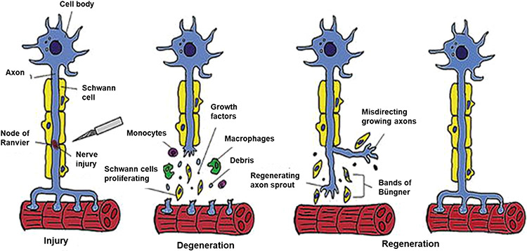
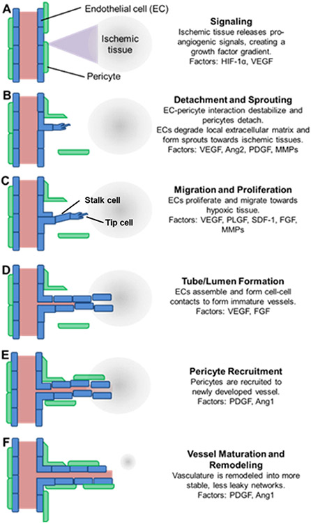
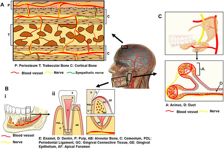
Craniofacial tissue injuries, diseases, and defects, including those within bone, dental, and periodontal tissues and salivary glands, impact an estimated 1 billion patients globally. Craniofacial tissue dysfunction significantly reduces quality of life, and successful repair of damaged tissues remains a significant challenge. Blood vessels and nerves are colocalized within craniofacial tissues and act synergistically during tissue regeneration. Therefore, the success of craniofacial regenerative approaches is predicated on successful recruitment, regeneration, or integration of both vascularization and innervation. Tissue engineering strategies have been widely used to encourage vascularization and, more recently, to improve innervation through host tissue recruitment or prevascularization/innervation of engineered tissues. However, current scaffold designs and cell or growth factor delivery approaches often fail to synergistically coordinate both vascularization and innervation to orchestrate successful tissue regeneration. Additionally, tissue engineering approaches are typically investigated separately for vascularization and innervation. Since both tissues act in concert to improve craniofacial tissue regeneration outcomes, a revised approach for development of engineered materials is required. This review aims to provide an overview of neurovascularization in craniofacial tissues and strategies to target either process thus far. Finally, key design principles are described for engineering approaches that will support both vascularization and innervation for successful craniofacial tissue regeneration.

Keywords: biomaterial design; cell therapy; craniofacial tissue; engineered tissue regeneration; growth factor; hydrogel; neurovascularization.


Figures

Figure 1.
Figure 1.
Depiction of Wallerian degeneration [Reproduced with permission from ref . Copyright 2017 IntechOpen].
Figure 2.
Figure 2.
Angiogenesis is a tightly controlled process involving numerous cells and factors. (A) Ischemic tissue increases production of hypoxia inducible factor 1α (HIF-1α), which induces production of vascular endothelial growth factor (VEGF) that (B) signals to pericytes (green) and endothelial cells (ECs, blue), resulting in detachment of pericytes and sprouting of endothelial tip cells toward the VEGF gradient. (C) Tip cells (blue) then migrate, degrade the matrix, and (D) enable stalk cells (blue) to proliferate and form vessels via alignment into tube-like luminal structures, followed by (E) pericyte recruitment and (F) further remodeling and maturation. HIF-1α, hypoxia inducible factor 1α; VEGF, vascular endothelial growth factor; Ang2, angiopoietin 2; PDGF, platelet derived growth factor; MMPs, matrix metalloproteinases; PLGF, placenta growth factor; SDF-1, stromal cell-derived factor 1; FGF, fibroblast growth factor; Ang1, angiopoietin 1 [Reproduced with permission from ref . Copyright 2015 Frontiers].
Figure 3.
Figure 3.
Anatomy of vascularized and innervated craniofacial tissues (A) cranial bone, (B) (i) mandibular bone (ii) and tooth, and (C) salivary glands. Part C was created with BioRender.com, agreement number: MB22XPLPSX.
Figure 4.
Figure 4.
(A) Longitudinal tracking of cranial bone defect healing in Col2.3GFP transgenic mice over a 6-week period illustrates osteogenesis and angiogenesis. Vessel, red; osteoblasts, green [Reproduced with permission from ref . Copyright 2018 Springer eBook]. (B) Reinnervation during cranial bone defect repair: representative whole-mount tile scans (left) and high-magnification images (right) of TUBB3 (beta III tubulin)-stained cranial defects (b–e) from days 3 to 28 postinjury compared to uninjured controls (a); TUBB3+ nerve fibers appear green; dashed white circles indicates margins of defect; white asterisks indicate midline suture and white scale bar, 200 mm [Reproduced with permission from ref . Copyright 2020 Elsevier].
Figure 5.
Figure 5.
(A) Representative confocal images of costained of CD31 (blood vessels, red) and β3-tubulin (nerves, green) on the cross sections of autografts, allografts, allografts modified by hydrolytically or MMP degradable tissue engineered periosteum (Hydro-TEP, MMP-TEP) at levels proximal (1), medial (2), and distal (3) in relation to the femoral head (scale bar = 20 μm) at 3-week postsurgery. (B) Blood vessel density-dependent effects on nerve density, where regression analysis demonstrates a linear relationship (R2 = 0.74) between revascularization and reinnervation, indicating their synergistic coordination during bone defect healing [Reproduced with permission from ref . Copyright 2021 Elsevier].
Figure 6.
Figure 6.
(A) In vivo release profiles of (i) VEGF and (ii) BMP-2 were obtained after implantation of scaffold into the subcutaneous tissue mice for 28 days, where CSB and CSV represent nanocomposite fibrous scaffold (CS) loaded with BMP2 (B) and VEGF (V), respectively [Reproduced with permission from ref . Copyright 2018 Elsevier]. (B) Release kinetics of PLGA microspheres steady release of neurotrophic growth factors was detected for at least 60 days (i, BDNF) and 80 days (ii, GDNF) [Reproduced with the permission from ref . Copyright 2010 Springer Nature]. (C) Normalized in vivo release profile of BMP-2 from the four different implants in a rat subcutaneous implantation model, where Mps = PLGA microparticles loaded with BMP-2, and PPF = poly(propylene fumarate) [Reproduced with the permission from ref . Copyright 2008 Elsevier]. (D) Cumulative release of VEGF from the biohybrid scaffold with PLGA nanofibers, which displays a sustained release of VEGF over 28 days [Reproduced with the permission from ref . Copyright 2014 Elsevier].

References

    1. Warren SM; Fong KD; Chen CM; Loboa EG; Cowan CM; Lorenz HP; Longaker MT Tools and techniques for craniofacial tissue engineering. Tissue Eng. 2003, 9 (2), 187–200. - PubMed
    1. Petrovic V; Zivkovic P; Petrovic D; Stefanovic V Craniofacial bone tissue engineering. Oral surgery, oral medicine, oral pathology and oral radiology 2012, 114 (3), No. e1. - PubMed
    1. Zhang W; Yelick PC Craniofacial tissue engineering. Cold Spring Harbor Perspect. Med 2018, 8 (1), No. a025775. - PMC - PubMed
    1. Li J; Gsaxner C; Pepe A; Morais A; Alves V; von Campe G; Wallner J; Egger J Synthetic skull bone defects for automatic patient-specific craniofacial implant design. Sci. Data 2021, 8 (1), 1–8. - PMC - PubMed
    1. Ozdemir D Dental caries: the most common disease worldwide and preventive strategies. Int. J. Biol 2013, 5 (4), 55.

Publication types

LinkOut - more resources