

Reduced-Dose Deep Learning Reconstruction for Abdominal CT of Liver Metastases
- PMID: 35014900
- PMCID: PMC8962777
- DOI: 10.1148/radiol.211838
Reduced-Dose Deep Learning Reconstruction for Abdominal CT of Liver Metastases
Abstract
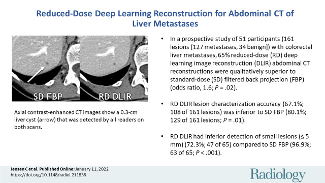
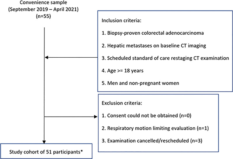
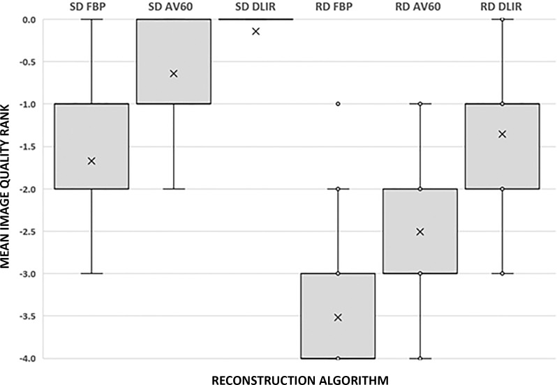
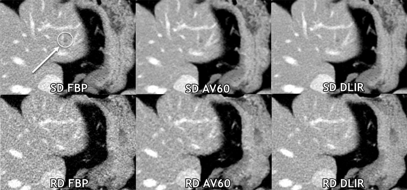
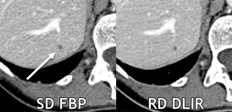
Background Assessment of liver lesions is constrained as CT radiation doses are lowered; evidence suggests deep learning reconstructions mitigate such effects. Purpose To evaluate liver metastases and image quality between reduced-dose deep learning image reconstruction (DLIR) and standard-dose filtered back projection (FBP) contrast-enhanced abdominal CT. Materials and Methods In this prospective Health Insurance Portability and Accountability Act-compliant study (September 2019 through April 2021), participants with biopsy-proven colorectal cancer and liver metastases at baseline CT underwent standard-dose and reduced-dose portal venous abdominal CT in the same breath hold. Three radiologists detected and characterized lesions at standard-dose FBP and reduced-dose DLIR, reported confidence, and scored image quality. Contrast-to-noise ratios for liver metastases were recorded. Summary statistics were reported, and a generalized linear mixed model was used. Results Fifty-one participants (mean age ± standard deviation, 57 years ± 13; 31 men) were evaluated. The mean volume CT dose index was 65.1% lower with reduced-dose CT (12.2 mGy) than with standard-dose CT (34.9 mGy). A total of 161 lesions (127 metastases, 34 benign lesions) with a mean size of 0.7 cm ± 0.3 were identified. Subjective image quality of reduced-dose DLIR was superior to that of standard-dose FBP (P < .001). The mean contrast-to-noise ratio for liver metastases of reduced-dose DLIR (3.9 ± 1.7) was higher than that of standard-dose FBP (3.5 ± 1.4) (P < .001). Differences in detection were identified only for lesions 0.5 cm or smaller: 63 of 65 lesions detected with standard-dose FBP (96.9%; 95% CI: 89.3, 99.6) and 47 lesions with reduced-dose DLIR (72.3%; 95% CI: 59.8, 82.7). Lesion accuracy with standard-dose FBP and reduced-dose DLIR was 80.1% (95% CI: 73.1, 86.0; 129 of 161 lesions) and 67.1% (95% CI: 59.3, 74.3; 108 of 161 lesions), respectively (P = .01). Lower lesion confidence was reported with a reduced dose (P < .001). Conclusion Deep learning image reconstruction (DLIR) improved CT image quality at 65% radiation dose reduction while preserving detection of liver lesions larger than 0.5 cm. Reduced-dose DLIR demonstrated overall inferior characterization of liver lesions and reader confidence. Clinical trial registration no. NCT03151564 © RSNA, 2022 Online supplemental material is available for this article.
Conflict of interest statement
Figures
References
-
- Solomon J , Marin D , Roy Choudhury K , Patel B , Samei E . Effect of radiation dose reduction and reconstruction algorithm on image noise, contrast, resolution, and detectability of subtle hypoattenuating liver lesions at multidetector CT: filtered back projection versus a commercial model-based iterative reconstruction algorithm . Radiology 2017. ; 284 ( 3 ): 777 – 787 . - PMC - PubMed
-
- Fält T , Söderberg M , Hörberg L , et al . Simulated dose reduction for abdominal CT with filtered back projection technique: effect on liver lesion detection and characterization . AJR Am J Roentgenol 2019. ; 212 ( 1 ): 84 – 93 . - PubMed
Publication types
MeSH terms
Associated data
Grants and funding
LinkOut - more resources
Full Text Sources
Medical
Miscellaneous
