. 2022 Jan 3;11(1):12.
doi: 10.1167/tvst.11.1.12.

Malondialdehyde-Modified Photoreceptor Outer Segments Promote Choroidal Neovascularization in Mice

Affiliations

Malondialdehyde-Modified Photoreceptor Outer Segments Promote Choroidal Neovascularization in Mice

Yuhong Chen et al. Transl Vis Sci Technol. .

Abstract

Purpose: This study aimed to establish a novel choroidal neovascularization (CNV) mouse model through subretinally injecting malondialdehyde (MDA)-modified photoreceptor outer segments (POS), which was more consistent with the pathogenesis of wet age-related macular degeneration (AMD).

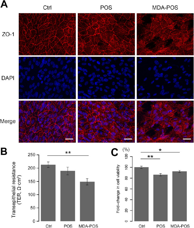
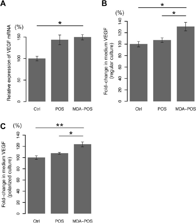
Methods: MDA-modified POS were subretinally injected in C57BL/6J mice. Four weeks later, to assess the volume of CNV and the morphology of retinal pigment epithelium (RPE), isolectin B4 and zonula occludens-1 antibody were used for immunostaining. Fundus fluorescent angiography and optical coherence tomography imaging were used to describe the morphologic features of CNV. Transepithelial resistance was measured on polarized ARPE-19 cells. Vascular endothelial growth factor levels in the cell culture medium were detected by enzyme-linked immunosorbent assay. The protein and messenger RNA expression levels of autophagy markers were measured using Western blot and quantitative polymerase chain reaction.

Results: CNV and RPE atrophy were successfully induced in the mouse model. MDA-modified POS also significantly increased the expression of vascular endothelial growth factor and disrupted cell junctions in RPE cells. In addition, MDA-modified POS induced autophagy-lysosomal impairment in RPE cells.

Conclusions: Subretinal injection of MDA-modified POS may generate a feasible CNV model that simulates the AMD pathological process.

Translational relevance: This study expands the understanding of the role of MDA in AMD pathogenesis, which provides a potential therapeutic target of AMD.


Conflict of interest statement

Disclosure: Y. Chen, None; X. Zhu, None; F. Ye, None; H. Wang, None; X. Wan, None; T. Zhang, None; Y. Wang, None; Y. Wang, None; X. Zhao, None; X. Bai, None; Y. Xiao, None; X. Sun, None

Figures

Figure 1.
Figure 1.
Subretinal injection of MDA-modified POS induced CNV in mice. (A) Fluorescein isothiocyanate-conjugated iB4 staining showed no CNV in any of the eyes in the control (PBS) group, but CNV was found in 4 of 10 eyes in the unmodified POS group, and 7 of 10 eyes in the MDA-modified POS group; the differences among the three groups were significant. (B–D) Representative flat-mounts of mouse choroids showed that the CNV volume in the unmodified POS group was smaller than that in the MDA-modified POS group (E and F) FFA at 4 weeks after subretinal injection. In the MDA-modified POS injection group, there was a hyperfluorescent spot. (G and H) Representative OCT scans at 4 weeks after subretinal injection. In the MDA-modified POS group, OCT images showed massive hyperreflectivity under the RPE layer, hyperreflective bands (arrowheads) at the border of the hyperreflective tissue, thinning outer nuclear layer (ONL), and disrupted photoreceptor layer. However, an intact and smooth RPE layer was observed in the control group. Scale bar, 50 µm. Ctrl, control. *P < 0.05 by t test, **P < 0.01 by Fisher's exact test.
Figure 2.
Figure 2.
Subretinal injection of MDA-modified POS induced RPE disruption in mice. Four weeks after subretinal injection, mouse choroid flat-mounts were obtained. ZO-1 staining showed that compared with unmodified POS, MDA-modified POS disrupted RPE tight junctions to a greater extent and increased the CNV volume. Subretinal injection of PBS caused almost no damage to the RPE. The top row of pictures represent a higher magnification of initial pictures. Scale bar, 100 µm.
Figure 3.
Figure 3.
MDA-modified POS disrupted the cell junctions of RPE cells under polarized culture conditions. (A) ARPE-19 cells were cultured under polarized conditions for eight weeks. ZO-1 staining showed that POS disrupted RPE tight junctions, and MDA-modified POS caused greater destruction than unmodified POS. (B) MDA-modified POS treatment significantly decreased the TER compared with that in the control group, which indicated that the cell-cell junctions had been functionally damaged. (C) POS treatment suppressed the viability of RPE cells compared with that of the control cells, but MDA modification did not further inhibit cell viability. Data are shown as the mean ± standard error of the mean. *P < 0.05, **P < 0.01 by the Kruskal–Wallis test.
Figure 4.
Figure 4.
MDA-modified POS induced VEGF expression. (A) Real-time polymerase chain reaction showed that VEGF messenger RNA in ARPE-19 cells under regular culture conditions was elevated by POS treatment, while the difference in VEGF messenger RNA levels between the unmodified and MDA-modified POS groups was not significant. VEGF in RPE cells grown under not only regular culture conditions (B) but also polarized culture conditions (C) was also detected by enzyme e-linked immunosorbent assay. VEGF secreted into the medium was increased in both groups compared with the control group. Data are shown as the mean ± standard error of the mean. *P < 0.05, **P < 0.01 by the Kruskal–Wallis test with the post hoc Steel–Dwass test.
Figure 5.
Figure 5.
MDA-modified POS induced changes in autophagy-related proteins in RPE cells. (A) Western blot analysis showed that the protein levels of Beclin-1 and LC3-II/LC3-I ratio were significantly increased and that the protein level of p62 was decreased after unmodified POS treatment compared with those in the control group. However, after treatment with MDA-modified POS, the protein expression of autophagy markers followed opposite trends. (B–D) The expression level of each protein was normalized to the mean value in the control group. Data are shown as the mean ± standard error of the mean. *P < 0.05, **P < 0.01 by the Kruskal–Wallis test with post hoc Steel–Dwass test.

References

    1. Mitchell P, Liew G, Gopinath B, et al. .. Age-related macular degeneration. Lancet. 2018; 392(10153): 1147–1159. - PubMed
    1. Niki E. Lipid peroxidation: physiological levels and dual biological effects. Free Radic Biol Med. 2009; 47(5): 469–484. - PubMed
    1. Uchida K. Aldehyde adducts generated during lipid peroxidation modification of proteins. Free Radic Res. 2015; 49(7): 896–904. - PubMed
    1. Njie-Mbye YF, Kulkarni-Chitnis M, Opere CA, et al. .. Lipid peroxidation: pathophysiological and pharmacological implications in the eye. Front Physiol. 2013; 4: 366. - PMC - PubMed
    1. Del Rio D, Stewart AJ, Pellegrini N.. A review of recent studies on malondialdehyde as toxic molecule and biological marker of oxidative stress. Nutr Metab Cardiovasc Dis. 2005; 15(4): 316–328. - PubMed

Publication types

Substances