. 2022 Jun;152(3):363-371.
doi: 10.1007/s11120-022-00896-w. Epub 2022 Jan 11.

Photosystem II antenna modules CP43 and CP47 do not form a stable 'no reaction centre complex' in the cyanobacterium Synechocystis sp. PCC 6803

Affiliations

Photosystem II antenna modules CP43 and CP47 do not form a stable 'no reaction centre complex' in the cyanobacterium Synechocystis sp. PCC 6803

Martina Bečková et al. Photosynth Res. 2022 Jun.

Abstract

The repair of photosystem II is a key mechanism that keeps the light reactions of oxygenic photosynthesis functional. During this process, the PSII central subunit D1 is replaced with a newly synthesized copy while the neighbouring CP43 antenna with adjacent small subunits (CP43 module) is transiently detached. When the D2 protein is also damaged, it is degraded together with D1 leaving both the CP43 module and the second PSII antenna module CP47 unassembled. In the cyanobacterium Synechocystis sp. PCC 6803, the released CP43 and CP47 modules have been recently suggested to form a so-called no reaction centre complex (NRC). However, the data supporting the presence of NRC can also be interpreted as a co-migration of CP43 and CP47 modules during electrophoresis and ultracentrifugation without forming a mutual complex. To address the existence of NRC, we analysed Synechocystis PSII mutants accumulating one or both unassembled antenna modules as well as Synechocystis wild-type cells stressed with high light. The obtained results were not compatible with the existence of a stable NRC since each unassembled module was present as a separate protein complex with a mutually similar electrophoretic mobility regardless of the presence of the second module. The non-existence of NRC was further supported by isolation of the His-tagged CP43 and CP47 modules from strains lacking either D1 or D2 and their migration patterns on native gels.

Keywords: CP43; CP47; No reaction centre complex; Photosynthesis; Photosystem II.


Conflict of interest statement

The authors have no conflict of interest to declare that are relevant to the content of this article.

Figures
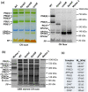
Fig. 1
Fig. 1
Analysis of membrane protein complexes of WT, ΔpsbC, ΔpsbB and ΔpsbE strains using CN electrophoresis on 4–14% polyacrylamide gel. a Isolated membranes were solubilized with n-β-dodecyl-maltoside and after electrophoresis, the gel was scanned for colour (CN scan) and chlorophyll fluorescence (CN fluor). Designation of complexes: PSI(3) and PSI(1), trimeric and monomeric photosystem I; PSII(2) and PSII(1), dimeric and monomeric Photosystem II; RC47, monomeric PSII lacking CP43; u.CP47’ and u.CP47, unassembled CP47 with and without bound Psb35, respectively; u.CP43 and u.CP43’, CP43 with and without PsbZ; and FP, free pigments. The samples were loaded on the same OD750nm basis corresponding to 4 µg of chlorophyll. b To visualize the markers, the gel in (a) was stained with Coomassie Brilliant Blue G-250 and scanned (CBB stained CN scan). Marker 1, high molecular weight calibration kit for electrophoresis (Amersham Biosciences); Marker 2, NativeMARK unstained protein standard (ThermoFisher). c The observed mass of protein complexes in gel (b) was calculated based on mobility of markers with known molecular mass
Fig. 2
Fig. 2
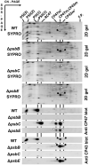
Analysis of membrane proteins of WT, ΔpsbC, ΔpsbB and ΔpsbE strains using 2D CN/SDS PAGE. Membrane complexes separated as shown in Fig. 1 were analysed using SDS PAGE in the second dimension and after analysis, the gels were stained with SYPRO Orange (SYPRO), electroblotted and CP43 and CP47 proteins were detected using specific antibodies. Designation of complexes as in Fig. 1, RC47 is monomeric PSII lacking CP43. Black and empty arrows designate CP43 and CP47 spots in the 2D-stained gels and blots, respectively. Loading was as in Fig. 1
Fig. 3
Fig. 3
Analysis of membrane proteins of WT treated with high light and cold stress. Cells of WT were treated with high light (300 µmol photons m−2 s−1) for 60 min at 29 °C (WT HL) or with high light (300 µmol photons m−2 s−1) for 60 min at 18 °C (WT HL/LT). Membranes isolated from these cells were solubilized with n-β-dodecyl-maltoside and after analysis on 6–14% polyacrylamide, CN gel was scanned for colour (1D colour) and chlorophyll fluorescence (1D fluor). Membrane complexes were then analysed using SDS PAGE in the second dimension and after analysis, the gel was stained with SYPRO Orange (2D SYPRO stain), electroblotted and CP47 and CP43 were detected using specific antibodies (2D blot). Designation of complexes and proteins as in Figs. 1 and 2. 4 µg of chlorophyll was loaded for each membrane preparation
Fig. 4
Fig. 4
Analysis of purified His-tagged CP43 and CP47 assembly modules using CN native gels. a His-PSI, His-CP47 and His-CP43 complexes were purified using nickel affinity chromatography from his-psaF, his-psbHpsbHpsbEftsH2 and his-psbC/ΔpsbA mutant strains. Isolated proteins were separated on 4–14% clear native gel (Komenda 2012a, b) together with WT membranes solubilized with 1% n-β-dodecyl-maltoside (w/v). Designation of complexes as in Figs. 1, 2 and 3, PSI(1)/CP43m is the monomeric photosystem I with bound CP43m; b His-CP43 and His-CP47 modules were purified from his-psbCpsbA and his-psbHpsbHpsbEftsH2-mutant strains, respectively, and separated on the 4–16% NativePAGE gel (ThermoFisher) (~ 0.5 mg of chlorophyll for the sample) together with WT membranes solubilized with 1% n-β-dodecyl-maltoside (w/v). Designation of complexes as in (a), CP47(2) refers to a dimer of CP47 module

Similar articles

Cited by

References

    1. Adir N, Shochat S, Ohad I. Light-dependent D1 protein synthesis and translocation is regulated by reaction center II. Reaction center II serves as an acceptor for the D1 precursor. J Biol Chem. 1990;265:12563–12568. doi: 10.1016/S0021-9258(19)38381-4. - DOI - PubMed
    1. Adir N, Zer H, Shochat S, et al. Photoinhibition – a historical perspective. Photosynth Res. 2003;76:343–370. doi: 10.1023/A:1024969518145. - DOI - PubMed
    1. Barbato R, Friso R, Rigoni F, et al. Structural changes and lateral redistribution of photosystem II during donor side photoinhibition of thylakoids. J Cell Biol. 1992;119:325–335. doi: 10.1083/jcb.119.2.325. - DOI - PMC - PubMed
    1. Barber J. Molecular Basis of the Vulnerability of Photosystem II to Damage by Light. Austr J Plant Physiol. 1995;22:201–208. doi: 10.1071/PP9950201. - DOI
    1. Boehm M, Romero E, Reisinger V, et al. Investigating the early stages of photosystem II assembly in Synechocystis sp. PCC 6803 isolation of CP47 and CP43 complexes. J Biol Chem. 2011;286:14812–14819. doi: 10.1074/jbc.M110.207944. - DOI - PMC - PubMed

MeSH terms

LinkOut - more resources