. 2022 Mar;59(3):1693-1705.
doi: 10.1007/s12035-021-02686-2. Epub 2022 Jan 11.

piRNA/PIWI Protein Complex as a Potential Biomarker in Sporadic Amyotrophic Lateral Sclerosis

Affiliations

piRNA/PIWI Protein Complex as a Potential Biomarker in Sporadic Amyotrophic Lateral Sclerosis

Rehab F Abdelhamid et al. Mol Neurobiol. 2022 Mar.

Abstract

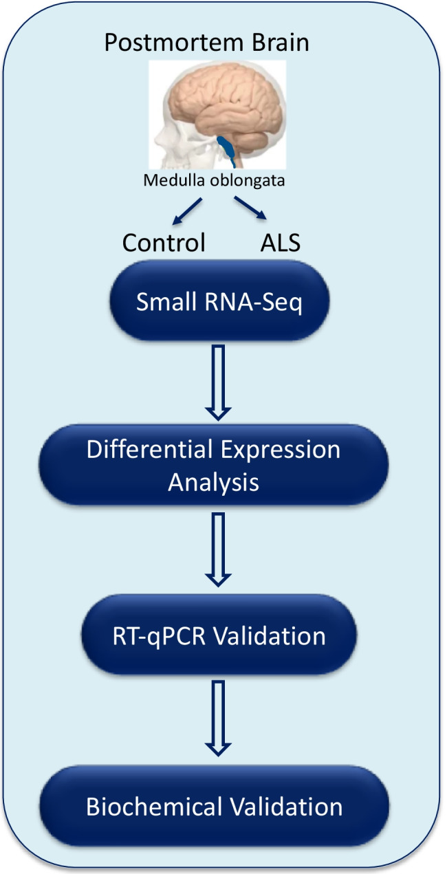
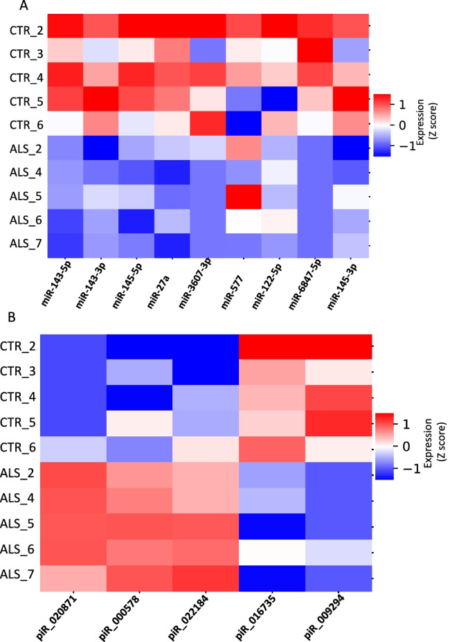
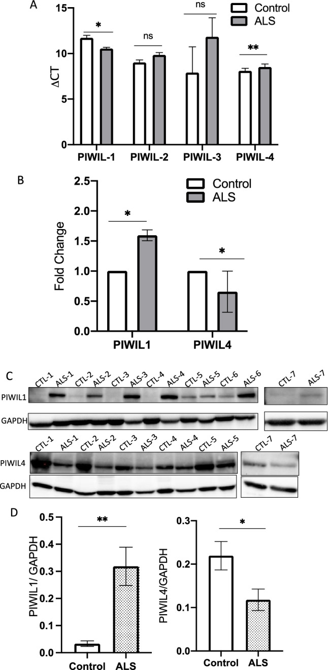
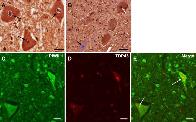
The pathological hallmark of the majority of amyotrophic lateral sclerosis (ALS) cases is the mislocalization and aggregation of TAR DNA-binding protein 43 (TDP-43), an RNA-binding protein. Several studies have attributed disease processes of ALS to abnormal RNA metabolism. However, dysregulated biogenesis of RNA, especially non-coding RNA (ncRNA), is poorly understood. To resolve it, RNA-Seq, biochemical, and immunohistochemical analyses were performed on the pyramidal tract of the medulla oblongata of sporadic ALS (sALS) and control postmortem brain samples. Here, we report perturbation of ncRNA biogenesis in PIWI-interacting RNA (piRNA) in several sALS brain samples associated with TDP-43 pathology. In addition, we confirmed the dysregulation of two PIWI homologs, PIWI-like-mediated gene silencing 1 (PIWIL1) and PIWIL4, which bind to piRNAs to regulate their expression. PIWIL1 was mislocalized and co-localized with TDP-43 in motor neurons of sporadic ALS lumbar cords. Our results imply that dysregulation of piRNA, PIWIL1, and PIWIL4 is linked to pathogenesis of ALS. Based on these results, piRNAs and PIWI proteins are potential diagnostic biomarkers and therapeutic targets of ALS.

Keywords: Amyotrophic lateral sclerosis; PIWI protein; TDP-43; miRNA; piRNA.


Conflict of interest statement

The authors declare no competing interests.

Figures

Fig. 1
Fig. 1
Schematic representation of the experimental design. Postmortem human tissue samples used in this study were collected from ALS cases (n = 7) and age- and sex-matched control cases (n = 7). RNA-Seq was performed on RNA extracted from the pyramidal tract of the medulla oblongata. Differential expression analysis of small RNAs was performed, followed by validation
Fig. 2
Fig. 2
miRNAs and piRNAs are dysregulated in sALS. A miRNAs are dysregulated in sALS. Differential expression analysis revealed that nine miRNAs were significantly dysregulated in ALS samples (p < 0.05). Data were normalized as the log 10 read count per million (RPM). B piRNAs are significantly dysregulated in sALS. Three piRNAs were up-regulated in ALS samples (red) and two were down-regulated (blue) (p < 0.05). Data were normalized as the log 10 read count per million (RPM)
Fig. 3
Fig. 3
Validation of piRNA expression with RT-qPCR. Dysregulation of five piRNAs was validated using RT-qPCR with two methods, the standard curve method to visualize the overall trend for piRNA perturbation (A) and the △CT method to quantify expression in individual samples (B). Both methods confirm that these five piRNAs were significantly altered in sALS samples in comparison with controls. A Average change in expression of each piRNA in control vs sALS samples. The Wilcoxon signed-rank test analysis was used for statistical calculations. Error bars denote SEMs. B piRNA expression by ΔCT in control vs sALS. The Mann–Whitney test was used for statistical calculations. ns: not significant. *p < 0.05, **p < 0.01
Fig. 4
Fig. 4
piRNA target genes are mostly ribosomal protein pseudogenes. The △CT method was used to investigate changes in expression of in silico-predicted target genes for dysregulated piRNA, according to piRBase and piRNAdb (Table 4). Except for TXRND1, target genes included ribosomal protein pseudogenes, RPL10P7, RPL13AP3, RPL18AP3, and 5S ribosomal RNA pseudogene transcripts, RNA5SP202 and RNA5-8SP6. We investigated the coding gene expression instead of pseudogenes due to increased evidence of ribosomal protein dysregulation in ALS. There was no notable change in gene expression except for expression of RPL13A (p = 0.053). This suggests that these piRNAs have regulatory effects beyond regulation of in silico-predicted target coding genes
Fig. 5
Fig. 5
PIWI proteins were dysregulated in postmortem sALS samples. A RT-qPCR for PIWIL1, PIWIL2, PIWIL3, PIWIL4 expression of mRNA using the ΔCT method. RT-qPCR using the △CT method for PIWIL1 and PIWIL4 showed significant differences between ALS and control samples, while PIWIL2 and PIWIL3 showed non-significant differences. B RT-qPCR results of postmortem PIWIL1 and PIWIL4 expression using a control pool for the standard curve method. One-tailed t-test and the Wilcoxon test were used for statistical calculations. PIWIL1 was upregulated 1.2–1.9-fold in sALS patients compared to the mean of control samples, by the standard curve method. C Western blot analysis of PIWIL1 and PIWIL4 in postmortem samples. The increase of PIWIL1 protein and the decrease of PIWIL4 protein were confirmed in ALS samples. D Quantitative analysis of PIWIL1 and PIWIL4 in a Western blot normalized to GAPDH. The increase of PIWIL1 protein and the decrease of PIWIL4 protein were statistically confirmed. ns: not significant. *p < 0.05, **p < 0.01
Fig. 6
Fig. 6
Immunohistochemistry for PIWIL1 in the lumbar spinal cord of control (A) and ALS (BE) patients. A In control patients, cell nuclei, the nuclear membrane (small arrows), and cell bodies of anterior horn cells (AHCs) are immunopositive for PIWIL1. B. In ALS patients, some remaining AHCs have nuclei positive for PIWIL1 (white arrow), while others have PIWIL1-negative nuclei (black arrow). CE. In double immunofluorescence staining, PIWIL1 is co-localized with intracytoplasmic inclusions positive for TDP-43 (white arrows) in ALS patients. Scale bar = 20 µm

References

    1. Mejzini R, Flynn LL, Pitout IL, Fletcher S, Wilton SD, Akkari PA, et al. ALS genetics, mechanisms, and therapeutics: where are we now? Front Neurosci. 2019;13:1310. doi: 10.3389/fnins.2019.01310. - DOI - PMC - PubMed
    1. Prasad A, Bharathi V, Sivalingam V, Girdhar A, Patel BK, et al. Molecular mechanisms of TDP-43 misfolding and pathology in amyotrophic lateral sclerosis. Front Mol Neurosci. 2019;12:25. doi: 10.3389/fnmol.2019.00025. - DOI - PMC - PubMed
    1. Wakisaka KT, Imai Y. The dawn of pirna research in various neuronal disorders. Front Biosci Landmark Ed. 2019;24:1440–1451. doi: 10.2741/4789. - DOI - PubMed
    1. Mao Q, Fan L, Wang X, Lin X, et al. Transcriptome-wide piRNA profiling in human brains for aging genetic factors. Jacobs J Genet. 2019;4:014. - PMC - PubMed
    1. Brettschneider J, Del Tredici K, Toledo JB, et al. Stages of pTDP-43 pathology in amyotrophic lateral sclerosis. Ann Neurol. 2013;74:20–38. doi: 10.1002/ana.23937. - DOI - PMC - PubMed

MeSH terms