Review
. 2022 Mar 2;110(5):749-769.
doi: 10.1016/j.neuron.2021.12.015. Epub 2022 Jan 10.

A functional subdivision within the somatosensory system and its implications for pain research

Affiliations
Review

A functional subdivision within the somatosensory system and its implications for pain research

Qiufu Ma. Neuron. .

Abstract

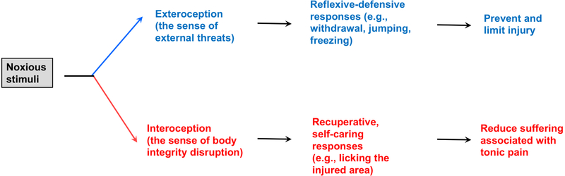
Somatosensory afferents are traditionally classified by soma size, myelination, and their response specificity to external and internal stimuli. Here, we propose the functional subdivision of the nociceptive somatosensory system into two branches. The exteroceptive branch detects external threats and drives reflexive-defensive reactions to prevent or limit injury. The interoceptive branch senses the disruption of body integrity, produces tonic pain with strong aversive emotional components, and drives self-caring responses toward to the injured region to reduce suffering. The central thesis behind this functional subdivision comes from a reflection on the dilemma faced by the pain research field, namely, the use of reflexive-defensive behaviors as surrogate assays for interoceptive tonic pain. The interpretation of these assays is now being challenged by the discovery of distinct but interwoven circuits that drive exteroceptive versus interoceptive types of behaviors, with the conflation of these two components contributing partially to the poor translation of therapies from preclinical studies.


Conflict of interest statement

Declaration of interests The author declares no competing interests.

Figures

Figure 1.
Figure 1.
Noxious stimuli evoke exteroceptive and interoceptive perceptions and associated behaviors.
Figure 2.
Figure 2.. Human studies reveal the segregation and convergence of the lateral versus medial thalamic pathways.
S1/S2: somatosensory cortex 1 and 2. ACC: anterior cingulate cortex. IC: insular cortex.
Figure 3:
Figure 3:. Molecular, developmental and functional classification of DRG neurons
Molecular classification (A) and developmental-functional segregation (B) of the sensory neurons in mouse dorsal root ganglia.
Figure 4.
Figure 4.. Partially segregated neural pathways driving different types of behaviors under acute conditions.
Adult Runx1-transient (“Runx1-trans.”; for example, TRPV1low-med) neurons (see Figure 3) may be connected to the spinal-slPBN-medial thalamic pathways that drive acute tonic pain and interoceptive self-caring behaviors such as persistent licking around the injured area. Adult Runx1-persistent neurons (see Figure 3) drive spinal reflexes vial local circuits and behavioral thermoregulation via the spinal-dlPBN-hypothalamic pathways plus the BLA pathway, although the route leading to BLA activation remains unknown (black “?”). Runx1-negative Aδ nociceptors also contribute to reflexive behaviors, such as reflexes in response to pinpricking stimulation by a thin needle (Arcourt et al., 2017; Qi et al., 2020). it remains unclear if defensive reactions such as freezing and jumping are driven by Runx1-persistent, Runx1-negative Aδ nociceptors (green dashed arrow) and/or Runx1-transient TRPV1low-medium neurons (red dashed arrow). PB: parabrachial nuclei. slPBN, dlPBN and elPBN: the superior lateral, the dorsolateral, and exterior lateral PB nuclei, respectively. BLA: the basal lateral nucleus of the amygdala. Hypothal.: hypothalamic nuclei. lPAG: the lateral periaqueductal gray nuclei. MdD: the dorsal reticular formation of the medullar (MdD). CeA: the central nuclei of the amygdala. Tac1-neg.: spinal Tac1-negative neurons.

References

    1. Abdel Samad O, Liu Y, Yang FC, Kramer I, Arber S, and Ma Q (2010). Characterization of two Runx1-dependent nociceptor differentiation programs necessary for inflammatory versus neuropathic pain. Mol Pain 6, 45. - PMC - PubMed
    1. Abdus-Saboor I, Fried NT, Lay M, Burdge J, Swanson K, Fischer R, Jones J, Dong P, Cai W, Guo X, et al. (2019). Development of a Mouse Pain Scale Using Sub-second Behavioral Mapping and Statistical Modeling. Cell Rep 28, 1623–1634 e1624. - PMC - PubMed
    1. Abraira VE, and Ginty DD (2013). The sensory neurons of touch. Neuron 79, 618–639. - PMC - PubMed
    1. Abraira VE, Kuehn ED, Chirila AM, Springel MW, Toliver AA, Zimmerman AL, Orefice LL, Boyle KA, Bai L, Song BJ, et al. (2017). The Cellular and Synaptic Architecture of the Mechanosensory Dorsal Horn. Cell 168, 295–310 e219. - PMC - PubMed
    1. Ackerley R, and Watkins RH (2018). Microneurography as a tool to study the function of individual C-fiber afferents in humans: responses from nociceptors, thermoreceptors, and mechanoreceptors. J Neurophysiol 120, 2834–2846. - PubMed

Publication types