Review
. 2022 Mar;142(3 Pt B):793-803.
doi: 10.1016/j.jid.2021.11.031. Epub 2022 Jan 10.

Type I Interferons in Autoimmunity

Affiliations
Review

Type I Interferons in Autoimmunity

Ruth Fernandez-Ruiz et al. J Invest Dermatol. 2022 Mar.

Abstract

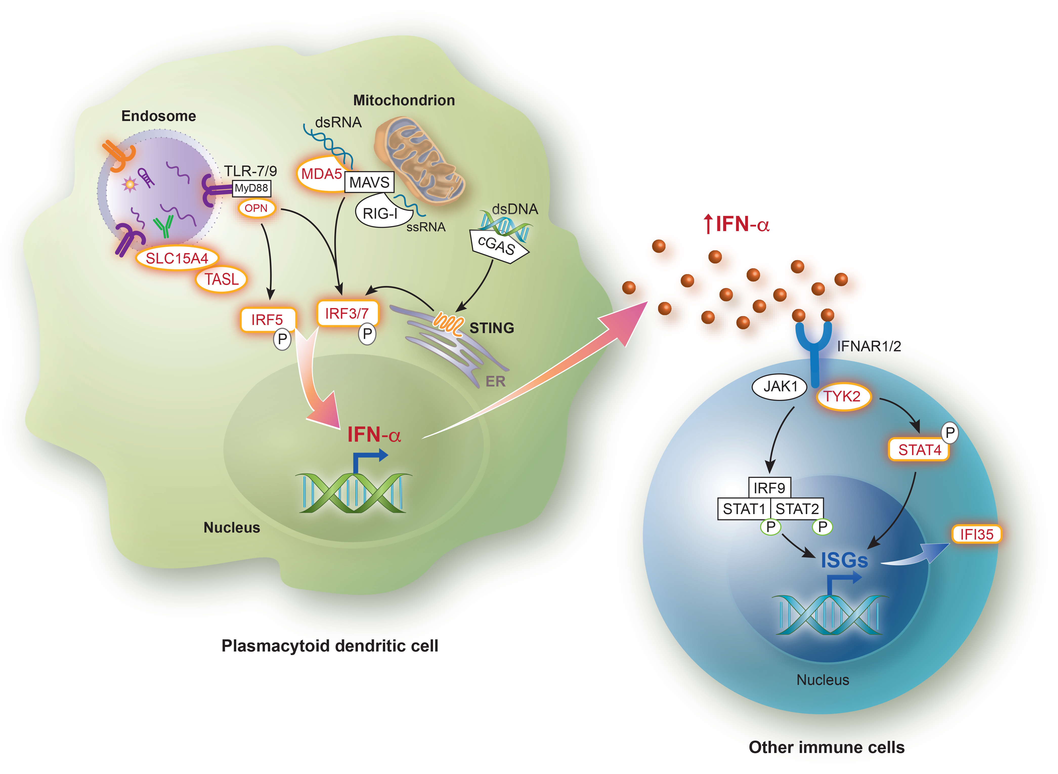
Dysregulated IFN-1 responses play crucial roles in the development of multiple forms of autoimmunity. Many patients with lupus, systemic sclerosis, Sjogren's syndrome, and dermatomyositis demonstrate enhanced IFN-1 signaling. IFN-1 excess is associated with disease severity and autoantibodies and could potentially predict response to newer therapies targeting IFN-1 pathways. In this review, we provide an overview of the signaling pathway and immune functions of IFN-1s in health and disease. We also review the systemic autoimmune diseases classically associated with IFN-1 upregulation and current therapeutic strategies targeting the IFN-1 system.


Figures

Figure 1.
Figure 1.. Risk alleles affecting type I interferon-related pathways in systemic autoimmune diseases.
Risk variants directly impacting the production of type I IFN or response that have been identified in SLE, Sjogren’s syndrome, systemic sclerosis, or dermatomyositis are shown in red. Immune complexes containing autoantibodies and self nucleic acids (or antibodies and viral particles) are endocytosed by plasmacytoid dendritic cells, leading to ligand-triggered activation of endosomal Toll-like receptors. The adaptor interacting with SLC15A4 on the Lysosome (TASL, encoded by the CXorf21 gene) is necessary for the recruitment and activation of IRF5 induced by ligand binding to the endosomal TLRs. TASL escapes chromosome X inactivation and is overexpressed in females compared to males, whereas SLC15A4 has been identified as an SLE risk loci. Following TLR-7 and TLR-9 activation, MyD88 and OPN are recruited, which allows for subsequent phosphorylation and nuclear translocation of IRF5 and/or IRF7. Polymorphisms in SPP1 (OPN), IRF5, IRF7 have also been identified in various type I IFN-related autoimmune diseases. Cytosolic nucleic acids are recognized by cGAS (double-stranded DNA), MDA5 (double-stranded RNA) or RIG-I (single-stranded RNA), after which the adaptor proteins MAVS (for MDA5 and RIG-I) and STING (for cGAS) are recruited, allowing for IRF3 phosphorylation and translocation to the nucleus. Activation of IFNAR by type I interferons, including IFN-α, results in phosphorylation of JAK1 and TYK2 and subsequent recruitment and phosphorylation of STAT proteins, which translocate to the nucleus and enhance expression of multiple interferon-stimulated genes (ISGs). TYK2 represents a susceptibility locus in SLE. STAT4 risk alleles have been identified in SLE and systemic sclerosis. The ISG IFI35, a negative regulator of the cytosolic nucleic acid sensor RIG-I, has been shown to be associated with dermatomyositis. cGAS, cyclic GMP–AMP synthase; dsDNA, double-stranded DNA; dsRNA, double-stranded RNA; IFI35, Interferon Induced Protein 35; IFN, Interferon; IFNAR, Interferon α/β receptor; IRF, Interferon regulatory factor; ISGs, Interferon-stimulated genes; JAK, Janus kinase; MAVS, mitochondrial antiviral-signaling protein; MDA5, Melanoma differentiation-associated protein 5; MyD88, myeloid differentiation factor 88; OPN, Osteopontin; RIG-I, Retinoic acid-inducible gene I; ssRNA; single-stranded RNA; STAT, Signal transducer and activator of transcription; STING, Stimulator of interferon genes; TASL, TLR adaptor interacting with SLC15A4 on the Lysosome; TLR, Toll-like receptor; TYK2, Tyrosine kinase 2. Illustration assistance provided by Jan Ruvido Stebbins, Ruvido Medical Illustration, Dexter, MI.

Similar articles

Cited by

References

    1. Absher DM, Li X, Waite LL, Gibson A, Roberts K, Edberg J, et al. Genome-wide DNA methylation analysis of systemic lupus erythematosus reveals persistent hypomethylation of interferon genes and compositional changes to CD4+ T-cell populations. PLoS Genet 2013;9(8):e1003678. - PMC - PubMed
    1. Ah Kioon MD, Tripodo C, Fernandez D, Kirou KA, Spiera RF, Crow MK, et al. Plasmacytoid dendritic cells promote systemic sclerosis with a key role for TLR8. Sci Transl Med 2018;10(423). - PMC - PubMed
    1. Alarcón-Riquelme ME, Ziegler JT, Molineros J, Howard TD, Moreno-Estrada A, Sánchez-Rodríguez E, et al. Genome-Wide Association Study in an Amerindian Ancestry Population Reveals Novel Systemic Lupus Erythematosus Risk Loci and the Role of European Admixture. Arthritis Rheumatol 2016;68(4):932–43. - PMC - PubMed
    1. Alarcón-Segovia D, Alarcón-Riquelme ME, Cardiel MH, Caeiro F, Massardo L, Villa AR, et al. Familial aggregation of systemic lupus erythematosus, rheumatoid arthritis, and other autoimmune diseases in 1,177 lupus patients from the GLADEL cohort. Arthritis Rheum 2005;52(4):1138–47. - PubMed
    1. Ali S, Mann-Nüttel R, Schulze A, Richter L, Alferink J, Scheu S. Sources of Type I Interferons in Infectious Immunity: Plasmacytoid Dendritic Cells Not Always in the Driver’s Seat. Front Immunol 2019;10:778. - PMC - PubMed

Publication types

Substances