Review
. 2022 Feb:342:241-279.
doi: 10.1016/j.jconrel.2022.01.008. Epub 2022 Jan 10.

Non-viral vectors for RNA delivery

Affiliations
Review

Non-viral vectors for RNA delivery

Yi Yan et al. J Control Release. 2022 Feb.

Abstract

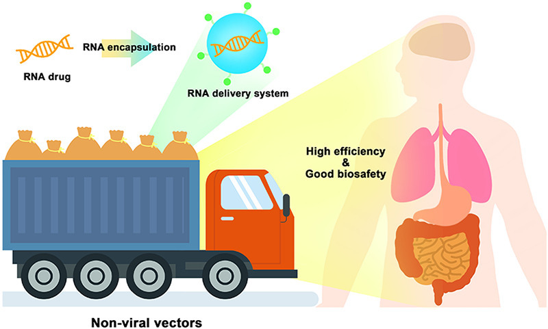
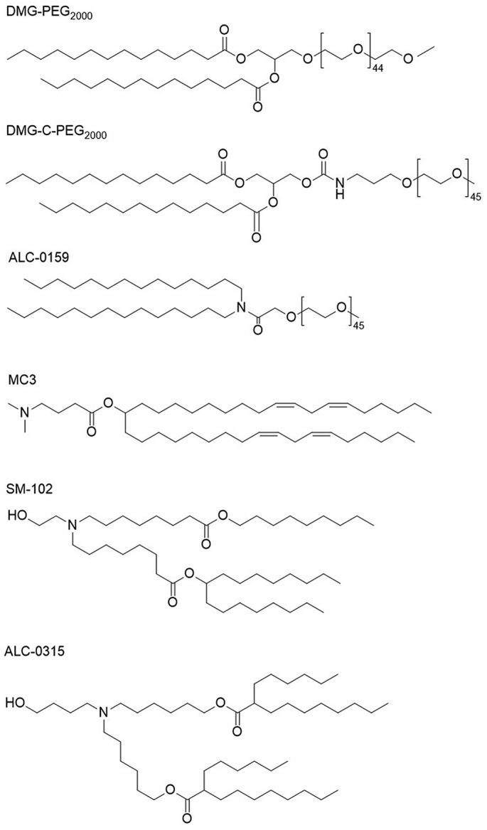
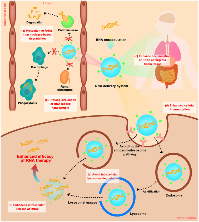
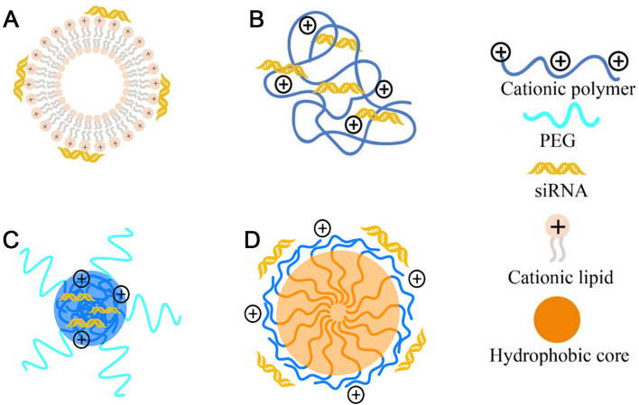
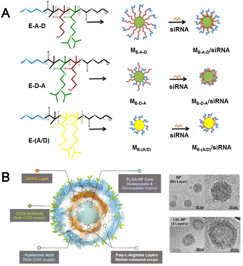
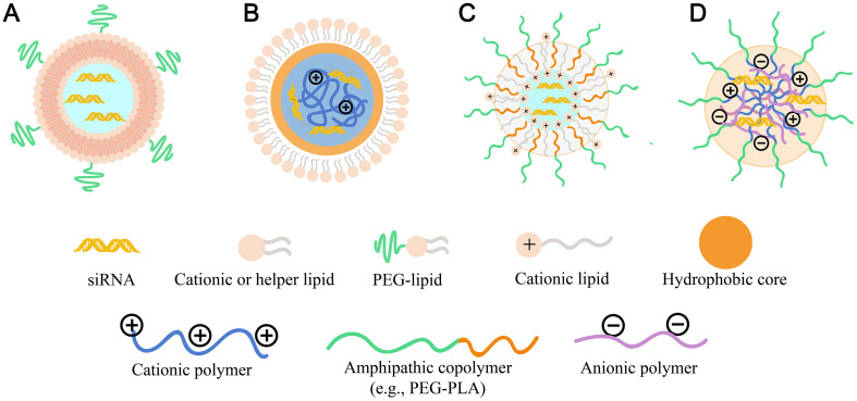
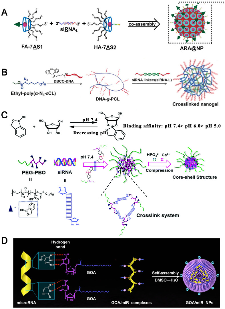
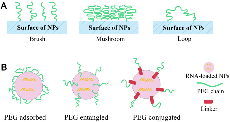
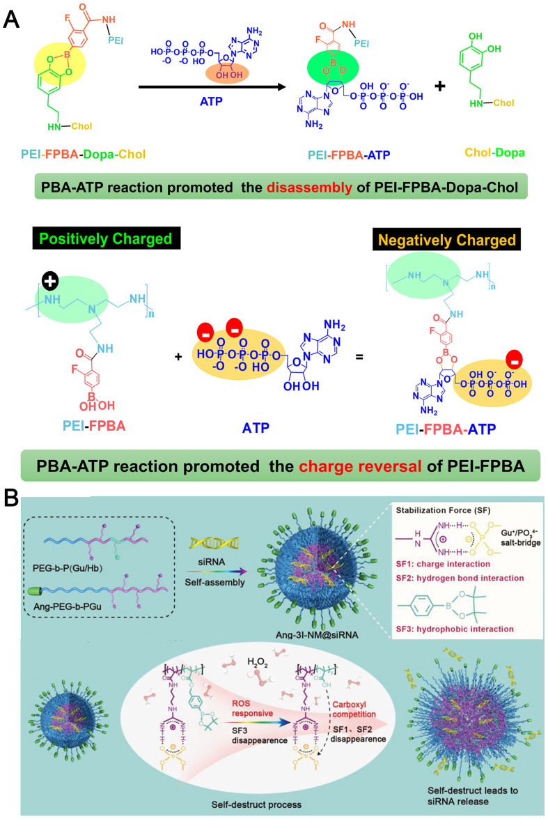
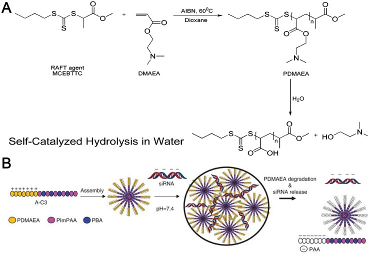
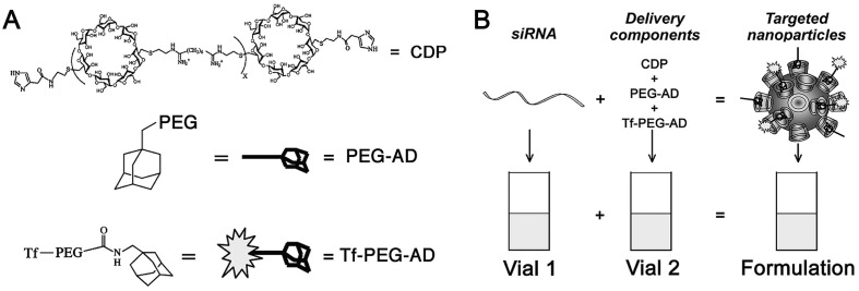
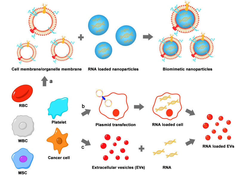
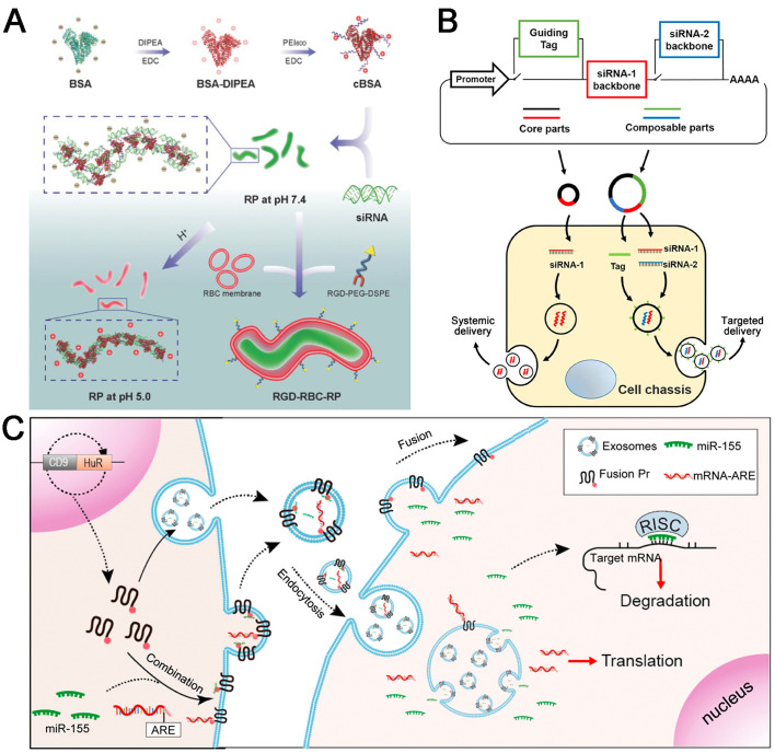
RNA-based therapy is a promising and potential strategy for disease treatment by introducing exogenous nucleic acids such as messenger RNA (mRNA), small interfering RNA (siRNA), microRNA (miRNA) or antisense oligonucleotides (ASO) to modulate gene expression in specific cells. It is exciting that mRNA encoding the spike protein of COVID-19 (coronavirus disease 2019) delivered by lipid nanoparticles (LNPs) exhibits the efficient protection of lungs infection against the virus. In this review, we introduce the biological barriers to RNA delivery in vivo and discuss recent advances in non-viral delivery systems, such as lipid-based nanoparticles, polymeric nanoparticles, N-acetylgalactosamine (GalNAc)-siRNA conjugate, and biomimetic nanovectors, which can protect RNAs against degradation by ribonucleases, accumulate in specific tissue, facilitate cell internalization, and allow for the controlled release of the encapsulated therapeutics.

Keywords: Biological barrier; Control release; Gene therapy; Non-viral vector; RNA drugs.


Conflict of interest statement

The authors stated that they have no competing interests.

Figures

Unlabelled Image
Graphical abstract
Fig. 1
Fig. 1
PEG-lipids and lonizable cationic lipids of Patisiran, BNT162b2 and mRNA-1273.
Fig. 2
Fig. 2
Extracellular and intracellular barriers for in vivo delivery of RNAs using non-viral vectors. (a) protection of RNAs from nuclease-based degradation; (b) prolong circulation of RNA-loaded nanocarriers by avoiding phagocytosis by mononuclear phagocytic system and rapid kidney clearance; (c) enhance tissue/organ-selective accumulation of RNAs; (d) enhance cellular internalization; (e) avoid intracellular lysosomal degradation; (f) enhance intracellular release of RNAs.
Fig. 3
Fig. 3
Protection of RNAs from nuclease degradation by electrostatic adsorption. The common cationic nanovectors for RNAs delivery including (A) cationic lipids-based lipoplexes, (B) cationic polymers-based polyplexes, (C) PEG-based cationic block copolymer formed NPs, and (D) cationic amphipathic-based block copolymer formed NPs.
Fig. 4
Fig. 4
(A) Molecular Structures of siRNA-loaded micelles of E–A–D, E–D–A, and E–(A/D) [95]. Reproduced with the permission from Ref. . Copyright © 2017 American Chemical Society. (B) Schematic illustration of a CD20/CD44 dual-targeted LbL-NP for precision siRNA therapeutics and Cryogenic TEM images of PLGA NPs (upper) and 4-layer LbL-NPs (down) [74]. Reproduced with the permission from Ref. . Copyright © 2019 John Wiley and Sons.
Fig. 5
Fig. 5
Protection of RNAs from nuclease degradation by electrostatic interaction-based layer-by-layer encapsulation and core-shell encapsulation. Illustration of (A) SNALP nanostructure; (B)lipid-polymer hybrid nanostructure (reverse micelle inner core); (C) polymer-lipid hybrid nanostructure (named as “CLAN”); and (D)PIC nanostructure.
Fig. 6
Fig. 6
(A) Schematic illustration of the co-assembly of branched antisense and siRNA for combined gene silencing and tumor therapy in vitro and in vivo. FA: folate for targeting; HA: influenza hemagglutinin peptide for endosomal escape; 7AS1 or 7AS2: branched antisenses covalently cross-linked by β-CD; siRNAL: 3′ terminal extended siRNA; and ARA@NP: FA-7AS1/siRNAL/HA-7AS2 co-assembled by hybridization between functionalized branched antisenses and siRNAL [105]. Reproduced with the permission from Ref. . Copyright © 2021 John Wiley and Sons. (B) Illustration of crosslinked nanogel formation and the siRNA delivery in vivo [106]. Reproduced with the permission from Ref. . Copyright © 2018 John Wiley and Sons. (C) Assembly of PEG-PBO/siRNA/CaP hybrid nanoparticles and its pH responsive disassembly [88]. Reproduced with the permission from Ref. . Copyright © 2018 Royal Society of Chemistry. (D) The nucleobase head of GOA prodrug was proposed to bind to nucleobase of miRNAs with hydrogen-bond interaction, and the oleic acid tail chains could provide hydrophobic forces to self-assemble into GOA/miR nanoparticles in aqueous solution [86]. Reproduced with the permission from Ref. . Copyright © 2019 Elsevier.
Fig. 7
Fig. 7
(A) Types of PEG conformations (B) techniques of linking PEG to NPs.
Fig. 8
Fig. 8
(A) siRNA polyplexes containing varied corona architectures. All polymers contain the same polyplex core-forming block consisting of equimolar DMAEMA and BMA. The corona-forming blocks comprise either linear PEG, zwitterionic PMPC, or brush PEG structures (POEGMA), as pictured. Polymer structures are displayed on the left, with the core-forming block in red and corona-forming block in blue. Polymers are complexed with siRNA at low pH, triggering spontaneous assembly of polyplexes before the pH is raised to physiological pH [120]. Reproduced with the permission from Ref . Copyright © 2017 American Chemical Society. (B) Schematic illustration of the adsorption efficacy of serum proteins onto the surface of aOEI-C12 NAs and f0.7OEI NAs [121]. Reproduced with the permission from Ref . Copyright © 2018 American Chemical Society.
Fig. 9
Fig. 9
(A) Addition of a supplemental component (termed a SORT molecule) to traditional LNPs systematically alters the in vivo delivery profile and mediates tissue-specific delivery as a function of the percentage and biophysical property of the SORT molecule [150]. Reproduced with the permission from Ref . Copyright © 2021 Springer Nature. (B) iPhos lipid design directs LNP tissue targeting. Zwitterion iPhos lipids contain two lipid tails on the positively charged tertiary amine and one lipid tail on the negatively charged phosphate group. Varying the alkyl lipid chain length impacts tissue targeting to liver, lung and spleen, which has been coined selective organ targeting (SORT) [151]. Reproduced with the permission from Ref . Copyright © 2021 Springer Nature. (C) Schematic representation of the formation of a disulfide bond between PDP-functionalized lipids that are incorporated into the liposome bilayer and reduced exofacial thiol groups, present at the cell surface [152]. Reproduced with the permission from Ref . Copyright © 2016 Elsevier.
Fig. 10
Fig. 10
(A) Synthesis of a siRNA-containing nanocaplet appended with transferrin (Tf) units (TfNC⊃siRNA) [158]. Reproduced with the permission from Ref. . Copyright © 2019 American Chemical Society. (B) Permeation of TfNC⊃siRNA into cells located in a deep tissue via Tf-mediated transcytosis. Once TfNC⊃siRNA escapes from the endosomes in a cell, glutathione (GSH), abundantly present in the cytoplasm, liberates siRNA to cause RNAi by reductive depolymerization of the nanocaplet part (TfNC) [158]. Reproduced with the permission from Ref. . Copyright © 2019 American Chemical Society. (C) Illustration of transmucus and transmembrane siTNF-α delivery mediated by fluorinated and guanidinated bifunctional helical polypeptides [159]. Reproduced with the permission from Ref. . Copyright © 2020 American Chemical Society.
Fig. 11
Fig. 11
(A) Scheme illustration showing the preparation of NPPLA/siRNA, NPPLGA/siRNA, and dPEGNPPLGA/siRNA with PEG5K-b-PLA11K, PEG5K-b-PLGA11K and the tumor pH-labile linkage-bridged block copolymer PEG5K-Dlinkm-PLGA11K. Compared with NPPLA/siRNA and NPPLGA/siRNA, dPEGNPPLGA/siRNA can enhance tumor cell uptake by detaching the PEG layer and accelerate intracellular siRNA release with hydrophobic PLGA layer [166]. Reproduced with the permission from Ref. . Copyright 2016 ELSEVIER. (B) Schematic illustration of constructing the Nb-modified nanogel (Nb-nanogel) [168]. Reproduced with the permission from Ref. . Copyright 2020 ELSEVIER.
Fig. 12
Fig. 12
(A) ATP-Triggered Breakage of FPBA-Dopa and ATP-Activated Charge Reversal of PEI-FPBA [196]. Reproduced with the permission from Ref. . Copyright © 2018 American Chemical Society. (B) Schematic illustration of formation of Ang-3I-NM@siRNA stabilized by the three “triple-interaction” forces, namely, electrostatic, hydrogen bond, and hydrophobic interactions, and mechanisms of ROS triggered siRNA release. In the presence of tumoral ROS, the hydrophobic phenylboronic ester is converted to the hydrophilic counterpart bearing carboxyl groups. This process first reduces the hydrophobic stabilization force and subsequently interferes with the electrostatic and hydrogen bond interactions resulting in effective siRNA release [195]. Reproduced with the permission from Ref. . Copyright © 2019 John Wiley and Sons.
Fig. 13
Fig. 13
(A) Polymerization and hydrolysis process of PDMAEA [203]. Reproduced with the permission from Ref. . Copyright 2011 American Chemical Society. (B) Mechanism for polymer assembly, binding with siRNA and release of siRNA through a self-catalyzed degradation of PDMAEA [198]. Reproduced with the permission from Ref. . Copyright 2013 Springer Nature.
Fig. 14
Fig. 14
Components and formulation of targeted NP-containing siRNA. (A) The delivery components are (i) a water-soluble, linear cyclodextrin-containing polymer (CDP), (ii) an adamantane (AD)-PEG conjugate (PEG MW of 5000) (AD-PEG), and (iii) the targeting component that is an adamantane conjugate of PEG (PEG MW of 5000) that has human transferrin (Tf) conjugated at the end opposite to the adamantane (Tf-PEG-AD). (B) The formulation contains two vials, one with siRNA and the other with the delivery components. When the two vials are mixed together, the targeted NPs form via self-assembly of the four components [213]. Reproduced with the permission from Ref. . Copyright © 2009 American Chemical Society.
Fig. 15
Fig. 15
Biomimetic vectors for RNAs delivery. Many kinds of cells can be used as a source for biomimetic vectors including RBC, white blood cell (WBC), platelet, mesenchymal stem cell (MSC), cancer cell, et al. cell membrane originated from these cells (a) can be directly used to coat RNA loaded nanoparticles to produce biomimetic nanoparticles inheriting donor cells' features. Or by transfecting plasmids into cells (b) to realize active RNA loading process with the help of endogenous mechanism of parent cells. Or by first isolating EVs and then loading RNA drugs (c) to construct RNA loaded EVs.
Fig. 16
Fig. 16
(A) Schematic illustration of preparation and charge-reversible profile of RGD-RBC-RP [16]. Reproduced with the permission from Ref. . Copyright © 2018 John Wiley and Sons. (B) Schematic description of the architecture of the genetic circuit [268]. Reproduced with the permission from Ref. . Copyright © 2021 Springer Nature. (C) Schematic summarization of the exosomes production by packaging cells via the RNA-HuR recognition [269]. Reproduced with the permission from Ref. . Copyright © 2019 American Chemical Society.

Similar articles

Cited by

References

    1. Anguela X.M., High K.A. Entering the modern era of gene therapy. Annu. Rev. Med. 2019;70:273–288. - PubMed
    1. Jeon Y.H., Jung Y.T. Production of a replicating retroviral vector expressing Reovirus fast protein for cancer gene therapy. J. Virol. Methods. 2022;299:114332. - PubMed
    1. Miname M.H., Rocha V.Z., Santos R.D. The role of RNA-targeted therapeutics to reduce ASCVD risk: what have we learned recently? Curr. Atheroscler. Rep. 2021;23(8):40. - PubMed
    1. https://www.fda.gov/news-events/press-announcements/fda-grants-accelerat...
    1. Kulkarni J.A., Witzigmann D., Thomson S.B., Chen S., Leavitt B.R., Cullis P.R., van der Meel R. Author correction: the current landscape of nucleic acid therapeutics. Nat. Nanotechnol. 2021;16(7):841. - PubMed

Publication types