. 2022 Aug;17(8):1776-1784.
doi: 10.4103/1673-5374.332156.

Essential role of MALAT1 in reducing traumatic brain injury

Affiliations

Essential role of MALAT1 in reducing traumatic brain injury

Na Wu et al. Neural Regen Res. 2022 Aug.

Abstract

As a highly evolutionary conserved long non-coding RNA, metastasis associated lung adenocarcinoma transcript 1 (MALAT1) was first demonstrated to be related to lung tumor metastasis by promoting angiogenesis. To investigate the role of MALAT1 in traumatic brain injury, we established mouse models of controlled cortical impact and cell models of oxygen-glucose deprivation to mimic traumatic brain injury in vitro and in vivo. The results revealed that MALAT1 silencing in vitro inhibited endothelial cell viability and tube formation but increased migration. In MALAT1-deficient mice, endothelial cell proliferation in the injured cortex, functional vessel density and cerebral blood flow were reduced. Bioinformatic analyses and RNA pull-down assays validated enhancer of zeste homolog 2 (EZH2) as a downstream factor of MALAT1 in endothelial cells. Jagged-1, the Notch homolog 1 (NOTCH1) agonist, reversed the MALAT1 deficiency-mediated impairment of angiogenesis. Taken together, our results suggest that MALAT1 controls the key processes of angiogenesis following traumatic brain injury in an EZH2/NOTCH1-dependent manner.

Keywords: EZH2; Jagged-1; LncRNA; MALAT1; NOTCH1; angiogenesis; controlled cortical impact; oxygen-glucose deprivation; traumatic brain injury; vascular remodeling.


Conflict of interest statement

None

Figures

Figure 1
Figure 1
MALAT1 expression profile and the effects of MALAT1 on neurological function. (A) Experimental design in vivo (A1) and in vitro (A2). (B) MALAT1 mRNA expression in brain tissue was detected by qRT-PCR at 6 hours and 1, 3, 7, 14, 21 and 28 days after CCI. The MALAT1 mRNA level rapidly increased as early as 6 hours after CCI, then gradually decreased to the lowest level at 7 days after CCI. (C) MALAT1 mRNA expression was detected at 24 hours after OGD by qRT-PCR. The MALAT1 level was increased in the OGD group compared with the sham group. (D) In situ hybridization detection of MALAT1 in vivo. MALAT1 (red, DyLight 594) co-localized with vWF (green, DyLight 488). Scale bars: 25 μm in D1–3, and 10 μm in D4–6. (E) The NSS. Zero points represented the minimum deficit, and ten points represented the maximum deficit. (F) The wire grip tests. Zero points represented the maximum deficit, and five points represented the minimum deficit. Data are shown as the mean ± SEM (n = 4/group in B–D, n = 6/group in E–F). *P < 0.05, **P < 0.01, ***P < 0.001 (one-way analysis of variance followed by Dunnett’s post hoc test in B, Student’s t-test in C or two-way repeated-measures analysis of variance followed by Tukey’s post hoc test in E–F). AAV: Adeno-associated virus; CBF: cerebral blood flow; CCI: controlled cortical impact; CCK8: Cell Counting Kit-8; EZH2: enhancer of zeste homolog 2; FISH: fluorescence in situ hybridization; GSK126: MALAT1 inhibitor; IF: immunofluorescence; Jagged-1: NOTCH1 signaling agonist; LNA: locked nucleic acid; LV: lentivirus; MALAT1: metastasis associated lung adenocarcinoma transcript 1; NSS: neurological severity score; OGD: oxygen-glucose deprivation; pcEZH2: recombinant plasmid pcDNA3.1(+)-EZH2 of overexpressing EZH2; PCR: polymerase chain reaction; qRT-PCR: quantitative reverse transcription polymerase chain reaction; siM: small interfering RNA-metastasis associated lung adenocarcinoma transcript 1; TBI: traumatic brain injury; vWF: von willebrand factor; WB: western blot.
Figure 2
Figure 2
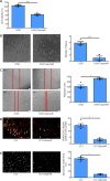
Effects of siRNA-mediated MALAT1 silencing on angiogenesis in vitro and in vivo. Cell viability (by Cell Counting Kit-8) (A), tube formation assay (B) and scratch wound assay (C) after OGD. (D–F) Effects of siMALAT1 on angiogenesis following CCI. The inhibition of MALAT1 suppressed endothelial proliferation and tube formation but promoted endothelial migration. (D) Double immunohistochemical staining of BrdU (green, DyLight 488, a marker of cell proliferation)/CD31 (red, DyLight 594, localized to positive endothelial cells) at 7 days following CCI. The number of BrdU/CD31 double-positive cells was significantly lower in the CCI-siM group compared with the CCI group. Arrows indicate the BrdU/CD31 double-positive cells. (E) Functional vessel density was identified by the staining of perfused lectin at 21 days following CCI. Functional vessel density was significantly lower in the CCI-siM group compared with the CCI group. Scale bars: 50 μm in B, 200 μm in C, 25 μm in D and 12.5 μm in E. (F) Effect of siMALAT1 on perfusion efficiency at the injury site 7 days following CCI, which was represented by two-dimensional laser speckle images of CBF changes (% CBF relative to contralateral hemisphere). CBF was significantly lower in the CCI-siM group compared with the CCI group. Circles indicate the CBF in lesion areas. Data are expressed as the mean ± SEM (n = 4/group in A–C, n = 5/group in D–F). *P < 0.05 (one-way analysis of variance followed by Tukey’s post hoc test). BrdU: Bromodeoxyuridine; CBF: cerebral blood flow; CCI: controlled cortical impact; MALAT1: metastasis associated lung adenocarcinoma transcript 1; OGD: oxygen-glucose deprivation; siM/siMALAT1: small interfering RNA-metastasis associated lung adenocarcinoma transcript 1.
Figure 3
Figure 3
siMALAT1 suppresses the NOTCH1 signaling pathway through EZH2 downregulation in vitro. (A) Bioinformatics prediction of MALAT1 and EZH2 interaction by StarBase. Left: Several candidate proteins potentially binding to MALAT1 listed in the StarBase system. Right: MALAT1 showed a high affinity to EZH2 in the RNA-protein interaction prediction system (random forest [RF] = 0.95 and support vector machine [SVM] scores = 0.76). (B) RNA pull-down assays confirmed the association between EZH2 and MALAT1. EZH2 was detected by western blot in collected samples pulled down by MALAT1. (C) EZH2 mRNA expression profile was detected by qRT-PCR following CCI at 6 hours and 1, 3, 7, 14, 21 and 28 days after CCI. (D) EZH2, NOTCH1 and HES1 expression levels were detected by western blot after MALAT1 downregulation in cultured endothelial cells. The expression levels of NOTCH1, HES1 and EZH2 were suppressed in the OGD-siM group compared with the OGD group. (E) Western blot was applied to test whether the NOTCH1 signaling pathway was regulated by an EZH2 inhibitor (GSK126) in cultured endothelial cells. (F) NOTCH1 and HES1 protein expression levels were detected by western blot after EZH2 overexpression combined with MALAT1 downregulation in cultured endothelial cells. Data were normalized to the sham group (C) or β-actin (D–F). Data are shown as the mean ± SEM (n = 4/group). *P < 0.05, **P < 0.01, ***P < 0.001 (one-way analysis of variance followed by Dunnett’s post hoc test in C, one-way analysis of variance followed by Tukey’s post hoc test in F, and Student’s t-test in B, D–E). CCI: Controlled cortical impact; EZH2: enhancer of zeste homolog 2; HES1: hairy and enhancer of split 1; MALAT1: metastasis associated lung adenocarcinoma transcript 1; OGD: oxygen-glucose deprivation; qRT-PCR: quantitative reverse transcription polymerase chain reaction; siM: small interfering RNA-metastasis associated lung adenocarcinoma transcript 1; TBI: traumatic brain injury.
Figure 4
Figure 4
MALAT1 regulates angiogenesis through the NOTCH1 signaling pathway in vitro and in vivo. Cell viability (by Cell Counting Kit-8) (A), tube formation assay (B) and scratch wound assay (C) after OGD. The effects of MALAT1 on endothelial proliferation, migration and tube formation after OGD stimuli were reversed by Jagged-1. (D) Double immunohistochemical staining of BrdU (green, DyLight 488, a marker of cell proliferation)/CD31 (red, DyLight 594, localized to positive endothelial cells) at 7 days following CCI. The number of BrdU/CD31 double-positive cells was significantly higher in the CCI-siM + Jagged-1 group compared with the CCI-siM group. Arrows indicate the BrdU/CD31 double-positive cells. (E) Functional vessel density was identified by the staining of perfused lectin at 21 days following CCI. Functional vessel density was significantly higher in the CCI-siM + Jagged-1 group compared with the CCI-siM group. Scale bars: 50 μm in B, 200 μm in C, 25 μm in D and 12.5 μm in E. (F) Effect of siMALAT1 combined with Jagged-1 on perfusion efficiency in injured sites 7 days following CCI. Two-dimensional laser speckle images of CBF changes (% CBF relative to contralateral hemisphere) in different groups following CCI. CBF was significantly higher in the CCI-siM + Jagged-1 group compared with the CCI-siM group. Circles indicate CBF in the lesion area. Data are shown as the mean ± SEM (n = 4/group in A–C, n = 5/group in D–F). *P < 0.05 (one-way analysis of variance followed by Tukey’s post hoc test). BrdU: Bromodeoxyuridine; CBF: cerebral blood flow; CCI: controlled cortical impact; Jagged-1: NOTCH1 signaling agonist; MALAT1: metastasis associated lung adenocarcinoma transcript 1; OGD: oxygen-glucose deprivation; siM/siMALAT1: small interfering RNA-metastasis associated lung adenocarcinoma transcript 1.
Figure 5
Figure 5
MALAT1 promotes neurological function through the NOTCH1 signaling pathway in vivo. (A) NSS: Higher points represent more serious deficits. (B) Wire grip tests: Higher points represent less serious deficits. Data are expressed as the mean ± SEM (n = 6/group). *P < 0.05 (two-way repeated-measures analysis of variance followed by Tukey’s post hoc test). CCI: Controlled cortical impact; NSS: neurological severity score; siM: small interfering RNA-metastasis associated lung adenocarcinoma transcript 1; TBI: traumatic brain injury.
Figure 6
Figure 6
Effects of LNA GapmeR-mediated inhibition of MALAT1 on angiogenesis in vitro and in vivo. Cell viability (by Cell Counting Kit-8) (A), tube formation assay (B) and scratch wound assay (C) after OGD. GapmeR suppressed angiogenic tube formation and endothelial proliferation and promoted endothelial migration. (D, E) Effects of GapmeR on angiogenesis following CCI. (D) Double immunohistochemical staining of BrdU (green, DyLight 488, a marker of cell proliferation)/CD31 (red, DyLight 594, localized to positive endothelial cells) at 7 days following CCI. The number of BrdU/CD31 double-positive cells was significantly lower in the CCI-GapmeR group compared with the CCI group. Arrows indicate the BrdU/CD31 double-positive cells. (E) Functional vessel density was identified by the staining of perfused lectin in different groups at 21 days following CCI. Functional vessel density was significantly lower in the CCI-GapmeR group compared with the CCI group. Scale bars: 50 μm in B, 200 μm in C, 25 μm in D and 12.5 μm in E. Data are shown as the mean ± SEM (n = 4/group in A–C, n = 5/group in D and E). *P < 0.05, ***P < 0.001 (Student’s t-test). BrdU: Bromodeoxyuridine; CCI: controlled cortical impact; LNA: locked nucleic acid; OGD: oxygen-glucose deprivation.

References

    1. Akil A, Gutiérrez-García AK, Guenter R, Rose JB, Beck AW, Chen H, Ren B. Notch signaling in vascular endothelial cells, angiogenesis, and tumor progression: an update and prospective. Front Cell Dev Biol. 2021;9:642352. - PMC - PubMed
    1. Asangani IA, Ateeq B, Cao Q, Dodson L, Pandhi M, Kunju LP, Mehra R, Lonigro RJ, Siddiqui J, Palanisamy N, Wu YM, Cao X, Kim JH, Zhao M, Qin ZS, Iyer MK, Maher CA, Kumar-Sinha C, Varambally S, Chinnaiyan AM. Characterization of the EZH2-MMSET histone methyltransferase regulatory axis in cancer. Mol Cell. 2013;49:80–93. - PMC - PubMed
    1. Beermann J, Piccoli MT, Viereck J, Thum T. Non-coding RNAs in development and disease: background, mechanisms, and therapeutic approaches. Physiol Rev. 2016;96:1297–1325. - PubMed
    1. Brütsch R, Liebler SS, Wüstehube J, Bartol A, Herberich SE, Adam MG, Telzerow A, Augustin HG, Fischer A. Integrin cytoplasmic domain-associated protein-1 attenuates sprouting angiogenesis. Circ Res. 2010;107:592–601. - PubMed
    1. Chen H, Xue LX, Cao HL, Chen SW, Guo Y, Gao WW, Ju SM, Tian HL. Endostatin/collagen XVIII is increased in cerebrospinal fluid after severe traumatic brain injury. Biomed Res Int. 2013;2013:402375. - PMC - PubMed