. 2022 Jan 11;13(1):199.
doi: 10.1038/s41467-021-27790-8.

A small RNA that cooperatively senses two stacked metabolites in one pocket for gene control

Affiliations

A small RNA that cooperatively senses two stacked metabolites in one pocket for gene control

Griffin M Schroeder et al. Nat Commun. .

Abstract

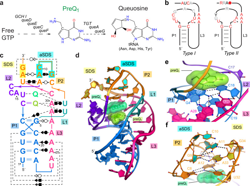
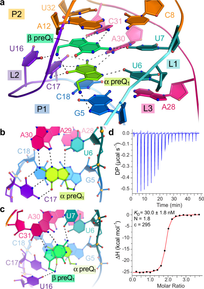
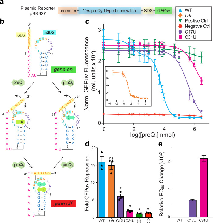
Riboswitches are structured non-coding RNAs often located upstream of essential genes in bacterial messenger RNAs. Such RNAs regulate expression of downstream genes by recognizing a specific cellular effector. Although nearly 50 riboswitch classes are known, only a handful recognize multiple effectors. Here, we report the 2.60-Å resolution co-crystal structure of a class I type I preQ1-sensing riboswitch that reveals two effectors stacked atop one another in a single binding pocket. These effectors bind with positive cooperativity in vitro and both molecules are necessary for gene regulation in bacterial cells. Stacked effector recognition appears to be a hallmark of the largest subgroup of preQ1 riboswitches, including those from pathogens such as Neisseria gonorrhoeae. We postulate that binding to stacked effectors arose in the RNA World to closely position two substrates for RNA-mediated catalysis. These findings expand known effector recognition capabilities of riboswitches and have implications for antimicrobial development.


Conflict of interest statement

The authors declare no competing interests.

Figures

Fig. 1
Fig. 1. Queuosine biosynthesis, the preQ1 riboswitch consensus model and co-crystal structure of the Carnobacterium antarcticus (Can) riboswitch.
a The queuosine (Q) biosynthetic pathway proceeds through the 7-deazapurine metabolite preQ1. b PreQ1-I riboswitch subtypes shown as secondary structures based on covariation. Red, black and gray positions indicate 97, 90, and 75% sequence conservation. Asterisk indicates a specificity base predicted to recognize preQ1. c Secondary structure of the Can riboswitch. Colors correspond to specific pseudoknot base pairing (P) and loop (L) sequences. PreQ1 is depicted as “Q”. Noncanonical pairing is indicated by Leontis–Westhof symbols. The Shine-Dalgarno sequence (SDS) and anti-(a)SDS are highlighted in yellow and cyan. d Ribbon diagram of the global Can riboswitch fold. e Binding pocket floor overview wherein floor bases comprise the A28•G5-C18 base triple. Dashed lines depict hydrogen bonds here and elsewhere. f Overview of the pocket ceiling, which comprises the U32•A12•C8 base triple. The view highlights P2 bases in the aSDS and SDS.
Fig. 2
Fig. 2. The Can preQ1-II riboswitch pocket with two preQ1 ligands and confirmation of ligand-to-RNA stoichiometry.
a Overview of fully occupied binding pocket. Interactions in the (b) α site and (c) β site. d Representative ITC experiment with titration of preQ1 into WT Can RNA. The binding constant KD, ligand-to-RNA stoichiometry N, and c value are shown.
Fig. 3
Fig. 3. Riboswitch reporter assay and dose response in live bacteria.
a Schematic of the plasmid reporter. b Two-site binding model wherein preQ1 can bind either site first. c Average GFPuv emission dependence on preQ1; (inset) one-site binding by the Lrh preQ1 riboswitch. d Bar graph showing fold repression of GFPuv emission for the Can, Lrh and mutant riboswitches with individual points shown. e Bar graph showing fold change in average EC50 relative to Can riboswitch EC50,2. Significance was determined by a two-tailed Student’s t test with Welch’s Correction (n = 3 biological replicates. *p ≤ 0.05). S.E.M. is shown in c and d; propagated errors are shown in e.

References

    1. Nahvi A, et al. Genetic control by a metabolite binding mRNA. Chem. Biol. 2002;9:1043–1049. - PubMed
    1. McCown PJ, et al. Riboswitch diversity and distribution. RNA. 2017;7:995–1011. - PMC - PubMed
    1. Breaker RR. Riboswitches and Translation Control. Cold Spring Harb. Perspect. Biol. 2018;10:1–14. - PMC - PubMed
    1. Suddala KC, et al. Single transcriptional and translational preQ1 riboswitches adopt similar pre-folded ensembles that follow distinct folding pathways into the same ligand-bound structure. Nucleic Acids Res. 2013;41:10462–10475. - PMC - PubMed
    1. Hua B, et al. Real-time monitoring of single ZTP riboswitches reveals a complex and kinetically controlled decision landscape. Nat. Commun. 2020;11:4531–4531. - PMC - PubMed

Publication types

MeSH terms