. 2022 Jan 11;12(1):504.
doi: 10.1038/s41598-021-04517-9.

Endoplasmic reticulum stress and the unfolded protein response in skeletal muscle of subjects suffering from peritoneal sepsis

Affiliations

Endoplasmic reticulum stress and the unfolded protein response in skeletal muscle of subjects suffering from peritoneal sepsis

Uta Barbara Metzing et al. Sci Rep. .

Abstract

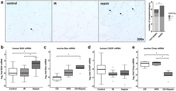
We provide a descriptive characterization of the unfolded protein response (UPR) in skeletal muscle of human patients with peritoneal sepsis and a sepsis model of C57BL/6J mice. Patients undergoing open surgery were included in a cross-sectional study and blood and skeletal muscle samples were taken. Key markers of the UPR and cluster of differentiation 68 (CD68) as surrogate of inflammatory injury were evaluated by real-time PCR and histochemical staining. CD68 mRNA increased with sepsis in skeletal muscle of patients and animals (p < 0.05). Mainly the inositol-requiring enzyme 1α branch of the UPR was upregulated as shown by elevated X-box binding-protein 1 (XBP1u) and its spliced isoform (XBP1s) mRNA (p < 0.05, respectively). Increased expression of Gadd34 indicated activation of PRKR-Like Endoplasmic Reticulum Kinase (PERK) branch of the UPR, and was only observed in mice (p < 0.001) but not human study subjects. Selected cell death signals were upregulated in human and murine muscle, demonstrated by increased bcl-2 associated X protein mRNA and TUNEL staining (p < 0.05). In conclusion we provide a first characterization of the UPR in skeletal muscle in human sepsis.


Conflict of interest statement

The authors declare no competing interests.

Figures

Figure 1
Figure 1
Markers of inflammation in skeletal muscle. Gene expression of inflammatory markers was assessed by RTqPCR in human (a) and murine (b) skeletal muscle. Data are given as normalized log2-fold changes compared to the median of the respective control groups as reference. Plots for human data represent n = 10 of matched subjects per group (for characterization see Suppl. Table 2), and plots for animal data represent n = 5–6 animals per group. Representative staining and semiquantitative assessment of CD68 in muscle tissue of patients (c) shows differences in number of macrophages (*p < 0.05, χ2-test). Arrows indicate exemplary CD68 positive cells (brownish coloring). CD control diet, CD/Cd68 cluster of differentiation 68, HFD high-fat diet, CCL chemokine C–C motif ligand, IR insulin resistance, MPO myeloperoxidase, RTqPCR quantitative real-time PCR, *p < 0.05.
Figure 2
Figure 2
Gene expression analysis of indicators of UPR activation in human (a) and murine (b) skeletal muscle. Data are given as normalized log2-fold changes compared to the median of the respective control groups as reference. Plots for human data represent n = 10 of matched subjects per group (Suppl. Table 2), and plots for animal data represent n = 5–6 animals per group. ATF activated transcription factor, BiP binding protein, also known as glucose-regulated protein 78 kDa; CD control diet, HFD high-fat diet, CD control diet, HFD high-fat diet, IR insulin resistance, XBP1s X-box protein 1 spliced, XBP1 X-box protein 1; *p < 0.05.
Figure 3
Figure 3
Indicators of apoptosis in skeletal muscle. TUNEL staining and semiquantitative assessment of positive cells (a) in human skeletal muscle in sepsis compared to the other groups (*p < 0.05, χ2-test). Arrows indicate TUNEL positive cells (brownish coloring), which are representative for cells undergoing apoptosis. Gene expression analysis for apoptotic marker genes BAX and CHOP, which are related to UPR activation, in human (b,d) and murine (c,e) skeletal muscle. Data are given as normalized log2-fold changes compared to the median of the respective control groups as reference. Plots for human data represent n = 10 of matched subjects per group (Suppl. Table 2), plots for animal data represent n = 5–6 animals per group. BAX/Bax bcl-2-associated X protein, CD control diet, CHOP/Chop C/EBP homologous protein, HFD high-fat diet, IR insulin resistance, TUNEL TdT-mediated dUTP-biotin nick end labeling; *p < 0.05.
Figure 4
Figure 4
Kaplan–Meier estimated curve of 28-day survival of patients suffering from peritoneal sepsis based on CD68 immunostaining in human skeletal muscle. 28-day survival was lower in patients with high number of infiltrating CD68 positive cells but not statistically significant in log-rank test (p = 0.06). CD68, cluster of differentiation 68.

References

    1. Angus DC, van der Poll T. Severe sepsis and septic shock. N. Engl. J. Med. 2013;369:840–851. doi: 10.1056/NEJMra1208623. - DOI - PubMed
    1. Singer M, et al. The third international consensus definitions for sepsis and septic shock (Sepsis-3) JAMA. 2016;315:801–810. doi: 10.1001/jama.2016.0287. - DOI - PMC - PubMed
    1. Callahan LA, Supinski GS. Sepsis-induced myopathy. Crit. Care Med. 2009;37:S354–S367. doi: 10.1097/CCM.0b013e3181b6e439. - DOI - PMC - PubMed
    1. Wischmeyer PE. Are we creating survivors…or victims in critical care? Delivering targeted nutrition to improve outcomes. Curr. Opin. Crit. Care. 2016;22:279–284. doi: 10.1097/MCC.0000000000000332. - DOI - PubMed
    1. van den Berghe G, et al. Intensive insulin therapy in mixed medical/surgical intensive care units: Benefit versus harm. Diabetes. 2006;55:3151–3159. doi: 10.2337/db06-0855. - DOI - PubMed

Publication types

MeSH terms