

Fluorescence lifetime DNA-PAINT for multiplexed super-resolution imaging of cells
- PMID: 35017652
- PMCID: PMC8752799
- DOI: 10.1038/s42003-021-02976-4
Fluorescence lifetime DNA-PAINT for multiplexed super-resolution imaging of cells
Abstract
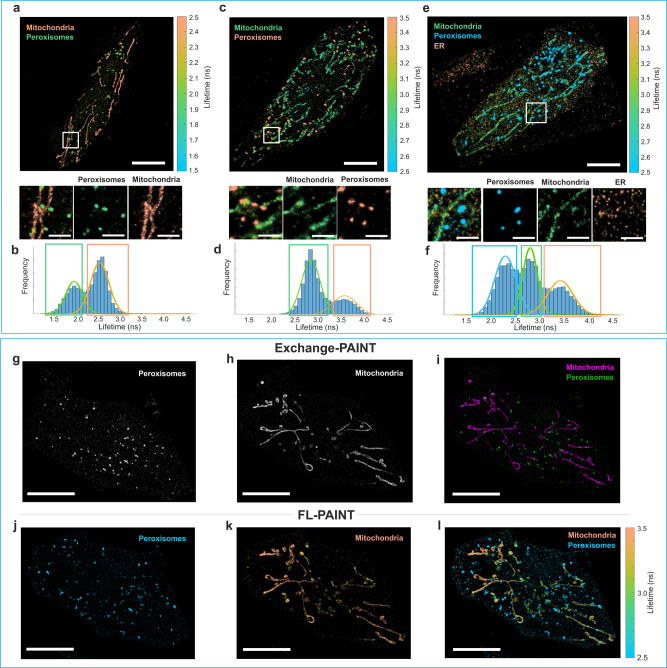
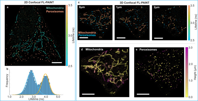
DNA point accumulation for imaging in nanoscale topography (DNA-PAINT) is a powerful super-resolution technique highly suitable for multi-target (multiplexing) bio-imaging. However, multiplexed imaging of cells is still challenging due to the dense and sticky environment inside a cell. Here, we combine fluorescence lifetime imaging microscopy (FLIM) with DNA-PAINT and use the lifetime information as a multiplexing parameter for targets identification. In contrast to Exchange-PAINT, fluorescence lifetime PAINT (FL-PAINT) can image multiple targets simultaneously and does not require any fluid exchange, thus leaving the sample undisturbed and making the use of flow chambers/microfluidic systems unnecessary. We demonstrate the potential of FL-PAINT by simultaneous imaging of up to three targets in a cell using both wide-field FLIM and 3D time-resolved confocal laser scanning microscopy (CLSM). FL-PAINT can be readily combined with other existing techniques of multiplexed imaging and is therefore a perfect candidate for high-throughput multi-target bio-imaging.
© 2022. The Author(s).
Conflict of interest statement
The authors declare no competing non-financial interests but the following competing financial interests: F.O. is a shareholder of NanoTag Biotechnologies GmbH. All other authors declare no competing interest.
Figures

Similar articles
-
Single-Molecule Fluorescence Lifetime Imaging Using Wide-Field and Confocal-Laser Scanning Microscopy: A Comparative Analysis.Nano Lett. 2022 Aug 10;22(15):6454-6461. doi: 10.1021/acs.nanolett.2c01586. Epub 2022 Jul 6. Nano Lett. 2022. PMID: 35792810 Free PMC article.
-
Fast, Background-Free DNA-PAINT Imaging Using FRET-Based Probes.Nano Lett. 2017 Oct 11;17(10):6428-6434. doi: 10.1021/acs.nanolett.7b03425. Epub 2017 Sep 21. Nano Lett. 2017. PMID: 28871786
-
Multiplexed 3D cellular super-resolution imaging with DNA-PAINT and Exchange-PAINT.Nat Methods. 2014 Mar;11(3):313-8. doi: 10.1038/nmeth.2835. Epub 2014 Feb 2. Nat Methods. 2014. PMID: 24487583 Free PMC article.
-
Advancements in DNA-PAINT: applications and challenges in biological imaging and nanoscale metrology.Nanoscale. 2025 Jun 12;17(23):14016-14034. doi: 10.1039/d4nr04544k. Nanoscale. 2025. PMID: 40407724 Review.
-
DNA-PAINT Super-Resolution Imaging for Characterization of Nucleic Acid Nanostructures.Chempluschem. 2022 Aug;87(8):e202200127. doi: 10.1002/cplu.202200127. Chempluschem. 2022. PMID: 35914775 Review.
Cited by
-
Super-Resolution Microscopy as a Versatile Tool in Probing Molecular Assembly.Int J Mol Sci. 2024 Oct 26;25(21):11497. doi: 10.3390/ijms252111497. Int J Mol Sci. 2024. PMID: 39519049 Free PMC article. Review.
-
Super-resolution imaging in whole cells and tissues via DNA-PAINT on a spinning disk confocal with optical photon reassignment.Nat Commun. 2025 May 29;16(1):4991. doi: 10.1038/s41467-025-60263-w. Nat Commun. 2025. PMID: 40442066 Free PMC article.
-
Super-resolution microscopy: a brief history and new avenues.Philos Trans A Math Phys Eng Sci. 2022 Apr 4;380(2220):20210110. doi: 10.1098/rsta.2021.0110. Epub 2022 Feb 14. Philos Trans A Math Phys Eng Sci. 2022. PMID: 35152764 Free PMC article.
-
Single-Molecule Fluorescence Lifetime Imaging Using Wide-Field and Confocal-Laser Scanning Microscopy: A Comparative Analysis.Nano Lett. 2022 Aug 10;22(15):6454-6461. doi: 10.1021/acs.nanolett.2c01586. Epub 2022 Jul 6. Nano Lett. 2022. PMID: 35792810 Free PMC article.
-
Fortunate molecules boost signal to background ratio and localization precision in correlation based single molecule localization microscopy.Commun Biol. 2024 Dec 23;7(1):1693. doi: 10.1038/s42003-024-07153-x. Commun Biol. 2024. PMID: 39715806 Free PMC article.
References
Publication types
MeSH terms
Substances
Grants and funding
LinkOut - more resources
Full Text Sources
Research Materials
