. 2022 Dec 29;118(16):3211-3224.
doi: 10.1093/cvr/cvac001.

Targeting RUNX1 as a novel treatment modality for pulmonary arterial hypertension

Affiliations

Targeting RUNX1 as a novel treatment modality for pulmonary arterial hypertension

Euy-Myoung Jeong et al. Cardiovasc Res. .

Abstract

Aims: Pulmonary arterial hypertension (PAH) is a fatal disease without a cure. Previously, we found that transcription factor RUNX1-dependent haematopoietic transformation of endothelial progenitor cells may contribute to the pathogenesis of PAH. However, the therapeutic potential of RUNX1 inhibition to reverse established PAH remains unknown. In the current study, we aimed to determine whether RUNX1 inhibition was sufficient to reverse Sugen/hypoxia (SuHx)-induced pulmonary hypertension (PH) in rats. We also aimed to demonstrate possible mechanisms involved.

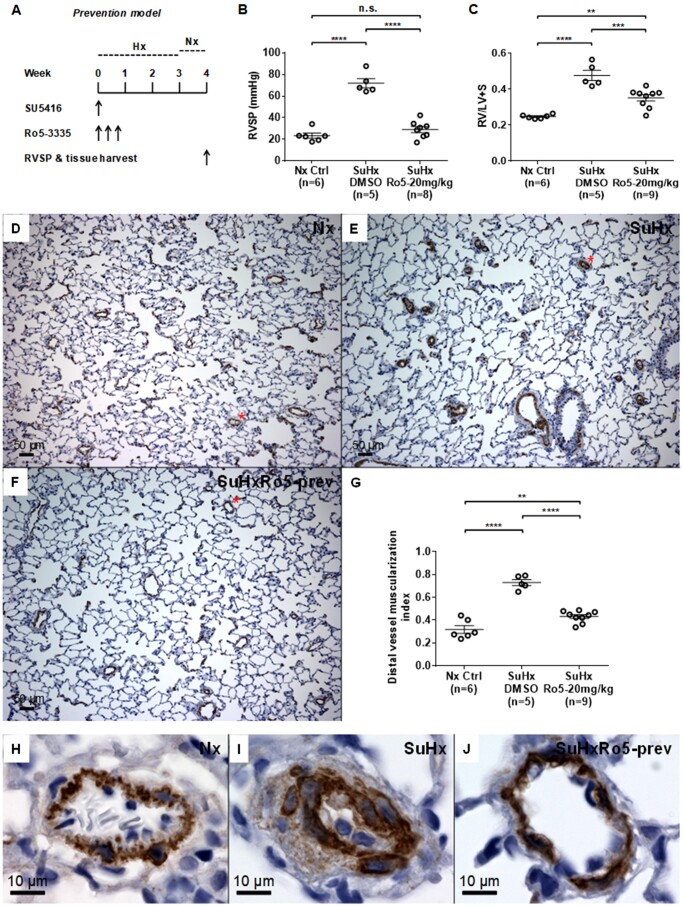
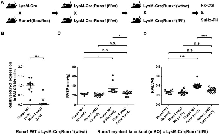
Methods and results: We administered a small molecule specific RUNX1 inhibitor Ro5-3335 before, during, and after the development of SuHx-PH in rats to investigate its therapeutic potential. We quantified lung macrophage recruitment and activation in vivo and in vitro in the presence or absence of the RUNX1 inhibitor. We generated conditional VE-cadherin-CreERT2; ZsGreen mice for labelling adult endothelium and lineage tracing in the SuHx-PH model. We also generated conditional Cdh5-CreERT2; Runx1(flox/flox) mice to delete Runx1 gene in adult endothelium and LysM-Cre; Runx1(flox/flox) mice to delete Runx1 gene in cells of myeloid lineage, and then subjected these mice to SuHx-PH induction. RUNX1 inhibition in vivo effectively prevented the development, blocked the progression, and reversed established SuHx-induced PH in rats. RUNX1 inhibition significantly dampened lung macrophage recruitment and activation. Furthermore, lineage tracing with the inducible VE-cadherin-CreERT2; ZsGreen mice demonstrated that a RUNX1-dependent endothelial to haematopoietic transformation occurred during the development of SuHx-PH. Finally, tissue-specific deletion of Runx1 gene either in adult endothelium or in cells of myeloid lineage prevented the mice from developing SuHx-PH, suggesting that RUNX1 is required for the development of PH.

Conclusion: By blocking RUNX1-dependent endothelial to haematopoietic transformation and pulmonary macrophage recruitment and activation, targeting RUNX1 may be as a novel treatment modality for pulmonary arterial hypertension.

Keywords: Endothelial cells; Macrophages; Pulmonary hypertension; RUNX1.


Conflict of interest statement

Conflict of interest: none declared.

Figures

Figure 1
Figure 1
Inhibition of RUNX1 in vivo prevents the development of Sugen/hypoxia-induced pulmonary hypertension (SuHx-PH) in rats. (A) Experimental protocol for prevention of SuHx-PH in rats shows administration of the RUNX1 inhibitor Ro5-3335 (Ro5) every other day for three times at the beginning of SuHx treatment. SU5416: VEGF receptor 2 antagonist Sugen 5416. (B and C) Right ventricular systolic pressure (RVSP) (B) and the Fulton’s index (right ventricle to left ventricle + septum, RV/LV+S ratio) (C) were measured 1 week after removal from 3 weeks of hypoxia (Hx). (DF) Representative microscopic images of 100× magnification show immunohistochemical (IHC) staining in brown colour of α-smooth muscle actin (α-SMA) in the blood vessels from lungs of normoxia control (Nx Ctrl) rats (D), vehicle DMSO-treated SuHx rats (E), and 20 mg/kg Ro5-treated SuHx rats (F). (G) Muscularization of distal pulmonary vessels less than 50 µm in diameter was assessed by calculating the muscularization index defined as the total area of the vessel that stained positive for α-SMA divided by total cross-sectional area of the vessel. (HJ) Representative small blood vessels stained in brown colour of α-SMA, which are labelled with a red asterisk in (DF), are shown in 600× magnification: (H) Nx Ctrl rats; (I) vehicle DMSO-treated SuHx rats; and (J) Ro5-treated SuHx rats. **P < 0.01, ***P < 0.001, ****P < 0.0001, n.s.: not significant, ordinary one-way ANOVA with multiple comparisons, n = number of animals in each experimental group.
Figure 2
Figure 2
Inhibition of RUNX1 in vivo reduces macrophage recruitment in the lung of SuHx-treated rats. (AC) Representative microscopic images of 100× magnification show IHC staining in dark brown colour of CD68+ macrophages in the lung of Nx rats (A), vehicle DMSO-treated SuHx rats (B), and Ro5-treated SuHx rats (C). (D) Three representative images of 100× magnification of the staining were taken for each sample and the number of CD68+ macrophages in each image was manually counted. A summary of the numbers of CD68+ macrophages per microscopic field is shown. (EG) Representative macrophages stained in dark brown colour for CD68, which are labelled with a red asterisk in (AC), are shown in 600× magnification: (E) Nx Ctrl rats; (F) vehicle DMSO-treated SuHx rats; and (G) Ro5-treated SuHx rats. ***P < 0.001, ****P < 0.0001, n.s.: not significant, ordinary one-way ANOVA with multiple comparisons, n = number of animals in each experimental group.
Figure 3
Figure 3
Inhibition of RUNX1 in vivo blocks the progression of SuHx-PH in rats. (A) Experimental protocol for intervention of SuHx-PH in rats shows administration of the RUNX1 inhibitor Ro5-3335 every other day for three times 1 week after the beginning of SuHx treatment. (B and C) RVSP (B) and RV/LV + S ratio (C) were measured 1 week after removal from 3 weeks of Hx. (DF) Representative microscopic images of 100× magnification show IHC staining in brown colour of α-SMA in the blood vessels from lungs of Nx Ctrl rats (D), vehicle DMSO-treated SuHx rats (E) and Ro5-treated SuHx rats (F). (G) Muscularization of distal pulmonary vessels less than 50 µm in diameter was assessed by calculating the muscularization index. (HJ) Representative small blood vessels stained in brown colour of α-SMA, which are labelled with a red asterisk in (DF), are shown in 600× magnification: (H) Nx Ctrl rats; (I) vehicle DMSO-treated SuHx rats; and (J) Ro5-treated SuHx rats. **P < 0.01, ***P < 0.001, ****P < 0.0001, n.s.: not significant, ordinary one-way ANOVA with multiple comparisons, n = number of animals in each experimental group.
Figure 4
Figure 4
Inhibition of RUNX1 in vivo reverses established SuHx-PH in rats. (A) Experimental protocol for reversal of SuHx-PH in rats shows administration of the RUNX1 inhibitor Ro5-3335 every other day for six times after the completion of SuHx treatment. (B and C) RVSP (B) and RV/LV + S ratio (C) were measured 2 weeks after removal from 3 weeks of Hx. (DF) Representative microscopic images of 100× magnification show IHC staining in brown colour of α-SMA in the blood vessels from lungs of Nx Ctrl rats (D), vehicle DMSO-treated SuHx rats (E) and Ro5-treated SuHx rats (F). (G) Muscularization of distal pulmonary vessels less than 50 µm in diameter was assessed by calculating the muscularization index. (HJ) Representative small blood vessels stained in brown colour of α-SMA, which are labelled with a red asterisk in (DF), are shown in 600× magnification: (H) Nx Ctrl rats; (I) vehicle DMSO-treated SuHx rats; and (J) Ro5-treated SuHx rats. *P < 0.05, ***P < 0.001, ****P < 0.0001, n.s.: not significant, ordinary one-way ANOVA with multiple comparisons, n = number of animals in each experimental group.
Figure 5
Figure 5
Genetic deletion of Runx1 in adult ECs/EPCs prevents SuHx-PH development in mice. (A) Experimental design to crossbreed Cdh5(PAC)-CreERT2 and Runx1(flox/flox) mice to generate Cdh5-CreERT; Runx1(fl/fl) mice, which were then subjected to tamoxifen treatment followed by SuHx-PH induction. (B) In 10- to 13-week-old mice, the loss of Runx1 in lung endothelial cells (ECs) upon Tam induction was verified per qRT–PCR. No changes in Runx1 gene expression in BM-derived CD14+ cells were found, demonstrating the endothelial specificity of Runx1 deletion in these mice. (C and D) Under Nx conditions, Cdh5-CreERT2; Runx1(wt/wt) mice, Cdh5-CreERT2; Runx1(fl/fl) mice treated with corn oil, and Cdh5-CreERT2; Runx1(fl/fl) mice treated with Tam all exhibited normal RVSP (C) and RV/LV + S ratio (D). Under SuHx conditions, Cdh5-CreERT2; Runx1(wt/wt) mice and Cdh5-CreERT2; Runx1(fl/fl) mice treated with corn oil exhibited significantly elevated RVSP (C) and RV/LV + S ratio (D). When the Cdh5-CreERT2; Runx1(fl/fl) mice were treated with Tam and placed under SuHx conditions, they exhibited normal RVSP (C) and RV/LV + S ratio (D). *P < 0.05, **P < 0.01, ***P < 0.001, ****P < 0.0001, n.s.: not significant, unpaired two-tailed Student’s t-test (B) and ordinary one-way ANOVA with multiple comparisons (C and D), n = number of animals in each experimental group.
Figure 6
Figure 6
Genetic deletion of Runx1 in cells of myeloid lineage prevents SuHx-PH development in mice. (A) Experimental design to crossbreed LysM-Cre and Runx1(flox/flox) mice to generate LysM-Cre; Runx1(fl/fl) mice, which were then subjected to SuHx-PH induction. (B) In 10- to 13-week-old mice, the loss of Runx1 in flow cytometry sorted BM-derived CD14+ monocytes was verified per qRT–PCR. (C and D) Under Nx conditions, LysM-Cre; Runx1(wt/wt) mice and LysM-Cre; Runx1(fl/fl) mice exhibited normal RVSP (C) and RV/LV + S ratio (D). Under SuHx conditions, LysM-Cre; Runx1(wt/wt) mice developed elevated RVSP (C) and RV/LV + S ratio (D), whereas the LysM-Cre; Runx1(fl/fl) mice maintained normal RVSP (C) and RV/LV + S ratio (D). *P < 0.05, ***P < 0.001, ****P < 0.0001, n.s.: not significant, unpaired two-tailed Student’s t-test (B) and ordinary one-way ANOVA with multiple comparisons (C and D), n = number of animals in each experimental group.
Figure 7
Figure 7
Tentative mechanisms with which targeting the haematopoietic transcription factor RUNX1 can be an effective novel treatment modality for PAH. Our model of pathogenesis of PAH (black arrows in clockwise) and proposed inhibition of RUNX1-dependent pathogenic processes indicated in red. Specific RUNX1 inhibition by a small molecule compound Ro5-3335 has the potential to block both the aberrant myeloid-skewed haematopoiesis and macrophage recruitment and activation, thus significantly impede disease development and progression.

References

    1. Farber HW, Loscalzo J.. Pulmonary arterial hypertension. N Engl J Med 2004;351:1655–1665. - PubMed
    1. Simonneau G, Galie N, Rubin LJ, Langleben D, Seeger W, Domenighetti G, Gibbs S, Lebrec D, Speich R, Beghetti M, Rich S, Fishman A.. Clinical classification of pulmonary hypertension. J Am Coll Cardiol 2004;43:5S–12S. - PubMed
    1. Simonneau G, Gatzoulis MA, Adatia I, Celermajer D, Denton C, Ghofrani A, Gomez Sanchez MA, Krishna Kumar R, Landzberg M, Machado RF, Olschewski H, Robbins IM, Souza R.. Updated clinical classification of pulmonary hypertension. J Am Coll Cardiol 2013;62:D34–D41. - PubMed
    1. McLaughlin VV, Shah SJ, Souza R, Humbert M.. Management of pulmonary arterial hypertension. J Am Coll Cardiol 2015;65:1976–1997. - PubMed
    1. Ventetuolo CE, Klinger JR.. WHO Group 1 pulmonary arterial hypertension: current and investigative therapies. Prog Cardiovasc Dis 2012;55:89–103. - PubMed

Publication types

MeSH terms

Substances