Review
. 2022 Apr;39(2):111-119.
doi: 10.1007/s10014-021-00423-0. Epub 2022 Jan 11.

A patient with two gliomas with independent oligodendroglioma and glioblastoma biology proved by DNA-methylation profiling: a case report and review of the literature

Affiliations
Review

A patient with two gliomas with independent oligodendroglioma and glioblastoma biology proved by DNA-methylation profiling: a case report and review of the literature

Theo F J Kraus et al. Brain Tumor Pathol. 2022 Apr.

Abstract

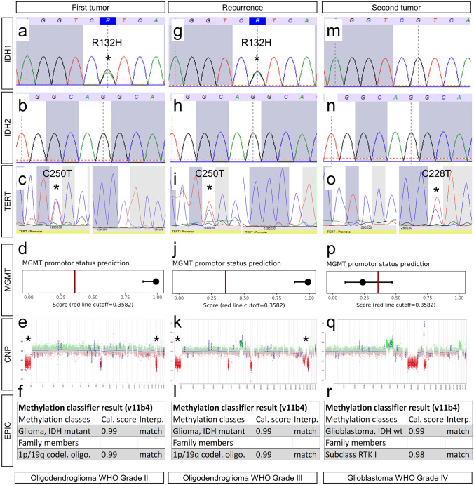
Here, we report on a patient presenting with two histopathologically distinct gliomas. At the age of 42, the patient underwent initial resection of a right temporal oligodendroglioma IDH mutated 1p/19q co-deleted WHO Grade II followed by adjuvant radiochemotherapy with temozolomide. 15 months after initial diagnosis, the patient showed right hemispheric tumor progression and an additional new left frontal contrast enhancement in the subsequent imaging. A re-resection of the right-sided tumor and resection of the left frontal tumor were conducted. Neuropathological work-up showed recurrence of the right-sided oligodendroglioma with features of an anaplastic oligodendroglioma WHO Grade III, but a glioblastoma WHO grade IV for the left frontal lesion. In depth molecular profiling revealed two independent brain tumors with distinct molecular profiles of anaplastic oligodendroglioma IDH mutated 1p/19q co-deleted WHO Grade III and glioblastoma IDH wildtype WHO grade IV. This unique and rare case of a patient with two independent brain tumors revealed by in-depth molecular work-up and epigenomic profiling emphasizes the importance of integrated work-up of brain tumors including methylome profiling for advanced patient care.

Keywords: Biomarker; DNA-methylation profiling; Glioblastoma; Glioma; Oligodendroglioma.


Conflict of interest statement

None declared.

Figures

Fig. 1
Fig. 1
Radiological findings over the course of the patients’ treatment. Axial postcontrast T1 weighted magnetic resonance imaging (MRI) showing a right-sided inhomogeneous contrast enhancing lesion located in the basal ganglia and peritrigonal area (a), as well as a parietal cystoid mass (b). After initial partial resection right peritrigonal tumor progression was seen (c and d). Furthermore, another left frontal rapidly progressive cystic tumor developed (e and f)
Fig. 2
Fig. 2
Histological and immunohistochemical findings. In H&E staining, the first tumor showed round shaped glial tumor cells with perinuclear halos (a). Immunohistochemistry with antibodies against GFAP showed positive tumor cells with only short processes (b). Reactions with antibodies against ATRX showed retained expression (c). Antibodies against IDH1 R132H mutant protein showed positive tumor cells (d). There were only sparse PHH3 positive cells (e). Proliferation was increased with 5% Ki67 positive cells (f). FISH analysis showed a combined loss of 1p (g) and 19q (h). Recurrence showed in H&E staining round tumor cells with perinuclear halos and brisk mitotic activity (i). Immunohistochemistry showed GFAP positive tumor cells (j). Nuclear expression of ATRX was retained (k). There was expression of IDH1 R132H mutant protein (l). There were increased PHH3 positive cells (m). Proliferation was increased with 25% Ki67 positive cells (n). FISH analysis of the 1p and 19q status revealed a combined loss of 1p (o) and 19q (p). Analysis of the second tumor showed in H&E staining a highly pleomorphic glia tumor with microvascular proliferation (q). Immunohistochemistry showed GFAP positive tumor cells (r). Nuclear expression of ATRX was retained (s). There was no expression of IDH1 R132H mutant protein (t). Reactions with antibodies against PHH3 showed increased mitoses (u). Proliferation was increased with 20% Ki67 positive cells (v). FISH analysis of the 1p and 19q status revealed no combined loss of 1p (w) and 19q (x)
Fig. 3
Fig. 3
Molecular genetic findings. The first tumor manifestation showed an IDH1 R132H mutation (a) with IDH2 wildtype (b) and TERT C250T promoter mutation (c). DNA Methylation profiling showed methylated MGMT promoter (d), 1p and 19q losses in CNP (e) and allocated the tumor to the methylation class of oligodendroglioma IDH mutated 1p/19q co-deleted (f). The recurrence showed IDH1 R132H mutation (g) with IDH2 wildtype (h) and TERT C250T promoter mutation (i). DNA-methylation profiling showed methylated MGMT promoter (j), 1p and 19q losses in CNP (k) and allocated the tumor to the methylation class of oligodendroglioma IDH mutated 1p/19q co-deleted (l). The second tumor showed IDH1 wildtype (m), IDH2 wildtype (n) and TERT C228T promoter mutation (o). DNA Methylation profiling showed unmethylated MGMT promoter (p), no 1p and 19q loss in CNP (q) and allocated the tumor to the methylation class of glioblastoma IDH wildtype, subclass RTK I (r). *: indication of IDH1 and TERT mutations and 1p/19q losses

References

    1. Capper D, Jones DTW, Sill M, et al. DNA methylation-based classification of central nervous system tumours. Nature. 2018;555:469–474. doi: 10.1038/nature26000. - DOI - PMC - PubMed
    1. Capper D, Stichel D, Sahm F, et al. Practical implementation of DNA methylation and copy-number-based CNS tumor diagnostics: the Heidelberg experience. Acta Neuropathol. 2018;136:181–210. doi: 10.1007/s00401-018-1879-y. - DOI - PMC - PubMed
    1. Halliday GC, Junckerstorff RC, Bentel JM, et al. The case for DNA methylation based molecular profiling to improve diagnostic accuracy for central nervous system embryonal tumors (not otherwise specified) in adults. J Clin Neurosci. 2018;47:163–167. doi: 10.1016/j.jocn.2017.09.013. - DOI - PubMed
    1. Holzl D, Hutarew G, Zellinger B, et al. Integrated analysis of programmed cell death ligand 1 expression reveals increased levels in high-grade glioma. J Cancer Res Clin Oncol. 2021;147:2271–2280. doi: 10.1007/s00432-021-03656-w. - DOI - PMC - PubMed
    1. Jaunmuktane Z, Capper D, Jones DTW, et al. Methylation array profiling of adult brain tumours: diagnostic outcomes in a large, single centre. Acta Neuropathol Commun. 2019;7:24. doi: 10.1186/s40478-019-0668-8. - DOI - PMC - PubMed