. 2022 Jan 10:116:e210176.
doi: 10.1590/0074-02760210176. eCollection 2022.

Identification and characterisation of SARS-CoV-2 and Human alphaherpesvirus 1 from a productive coinfection in a fatal COVID-19 case

Affiliations

Identification and characterisation of SARS-CoV-2 and Human alphaherpesvirus 1 from a productive coinfection in a fatal COVID-19 case

Alice Laschuk Herlinger et al. Mem Inst Oswaldo Cruz. .

Abstract

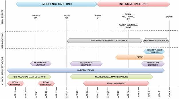
Background: During routine Coronavirus disease 2019 (COVID-19) diagnosis, an unusually high viral load was detected by reverse transcription real-time polymerase chain reaction (RT-qPCR) in a nasopharyngeal swab sample collected from a patient with respiratory and neurological symptoms who rapidly succumbed to the disease. Therefore we sought to characterise the infection.

Objectives: We aimed to determine and characterise the etiological agent responsible for the poor outcome.

Methods: Classical virological methods, such as plaque assay and plaque reduction neutralisation test combined with amplicon-based sequencing, as well as a viral metagenomic approach, were performed to characterise the etiological agents of the infection.

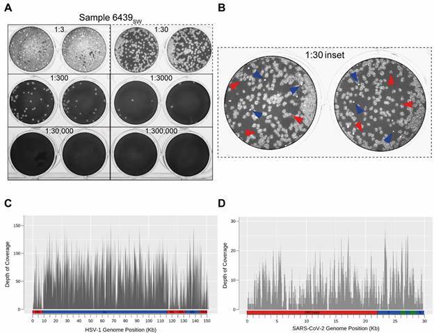
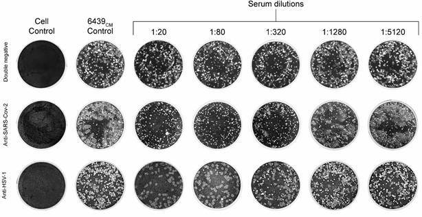
Findings: Plaque assay revealed two distinct plaque phenotypes, suggesting either the presence of two severe acute respiratory syndrome coronavirus 2 (SARS-CoV-2) strains or a productive coinfection of two different species of virus. Amplicon-based sequencing did not support the presence of any SARS-CoV-2 genetic variants that would explain the high viral load and suggested the presence of a single SARS-CoV-2 strain. Nonetheless, the viral metagenomic analysis revealed that Coronaviridae and Herpesviridae were the predominant virus families within the sample. This finding was confirmed by a plaque reduction neutralisation test and PCR.

Main conclusions: We characterised a productive coinfection of SARS-CoV-2 and Herpes simplex virus 1 (HSV-1) in a patient with severe symptoms that succumbed to the disease. Although we cannot establish the causal relationship between the coinfection and the severity of the clinical case, this work serves as a warning for future studies focused on the interplay between SARS-CoV-2 and HSV-1 coinfection and COVID-19 severity.


Figures

Fig. 1:
Fig. 1:. summarised timeline of the clinical evolution of patient 6439.
Fig. 2:
Fig. 2:. detection of viral coinfection of patient 6439. (A) Plaque assay of the nasopharyngeal swab sample from patient 6439 (6439SW) showing the indicated sample dilutions in duplicate wells. (B) The two different plaque phenotypes observed in this sample are highlighted in the inset of the 1:30 dilution duplicates (dashed panel). Red arrowheads correspond to the larger and turbid plaques with undefined borders, whereas blue arrowheads indicate the smaller and clearer plaques with well-defined circular borders. Depth of coverage diagram across the reference genome for Herpes simplex virus 1 (HSV-1) (C) and severe acute respiratory syndrome coronavirus 2 (SARS-CoV-2) (D) in kilobase (Kb) obtained through virome analysis. The basic structure of both genomes is indicated below the diagrams. SARS-CoV-2: non-structural coding region (red) indicated by ORF1a&b (the overlapping open reading frames 1a and 1b); and structural and accessory coding regions (blue and green) highlighting the S (spike) and N (nucleocapsid) genes. HSV-1: UL and US (blue) stand for Unique Long and Unique Short regions, respectively; and TR(L/S) and IR(L/S) (red) stand for Terminal and Internal - inverted - Repeats flanking the UL and the US regions, respectively.
Fig. 3:
Fig. 3:. characterisation of the two plaque phenotypes. Plaque reduction neutralisation test (PRNT) using double-negative, anti-severe acute respiratory syndrome coronavirus 2 (SARS-CoV-2), anti-Herpes simplex virus 1 (HSV-1) sera. Columns correspond to fixed Vero cell monolayers subjected to the following conditions from left to right: uninfected cells (cell control), 6439CM - inoculated cells without serum (virus control), and cells inoculated with 6439CM pre-incubated with the indicated dilutions of the corresponding serum.

References

    1. Matschke J, Lütgehetmann M, Hagel C, Sperhake JP, Schröder AS, Edler C. Neuropathology of patients with COVID-19 in Germany a post-mortem case series. Lancet Neurol. 2020;19:919–929. - PMC - PubMed
    1. Rabaan AA, Tirupathi R, Sule AA, Aldali J, Mutair AA, Alhumaid S. Viral dynamics and real-time RT-PCR Ct values correlation with disease severity in COVID-19. Diagnostics. 2021;11:1091–1091. - PMC - PubMed
    1. Voloch CM, Francisco RS, Jr, de Almeida LGP, Brustolini OJ, Cardoso CC, Gerber AL. Intra-host evolution during SARS-CoV-2 prolonged infection. Virus Evol. 2021;7(2):veab78–veab78. - PMC - PubMed
    1. Bolger AM, Lohse M, Usadel B. Trimmomatic a flexible trimmer for Illumina sequence data. Bioinformatics. 2014;30:2114–2120. - PMC - PubMed
    1. Martin M. Cutadapt removes adapter sequences from high-throughput sequencing reads. MBnet.journal [Internet] 2011;17:10–10.