. 2022 Apr;132(4):2746-2759.
doi: 10.1111/jam.15449. Epub 2022 Feb 11.

Membrane vesicles from antibiotic-resistant Staphylococcus aureus transfer antibiotic-resistance to antibiotic-susceptible Escherichia coli

Affiliations

Membrane vesicles from antibiotic-resistant Staphylococcus aureus transfer antibiotic-resistance to antibiotic-susceptible Escherichia coli

Ae Rin Lee et al. J Appl Microbiol. 2022 Apr.

Abstract

Aim: Bacteria naturally produce membrane vesicles (MVs), which have been shown to contribute to the spread of multi-drug resistant bacteria (MDR) by delivering antibiotic-resistant substances to antibiotic-susceptible bacteria. Here, we aim to show that MVs from Gram-positive bacteria are capable of transferring β-lactam antibiotic-resistant substances to antibiotic-sensitive Gram-negative bacteria.

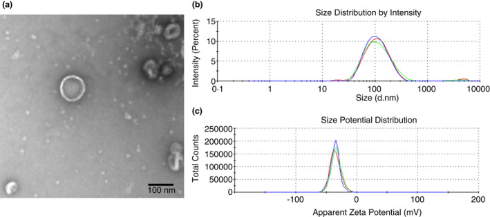
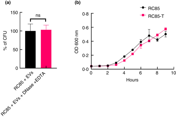
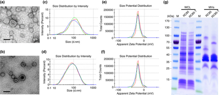
Materials and methods: MVs were collected from a methicillin-resistant strain of Staphylococcus aureus (MRSA) and vesicle-mediated fusion with antimicrobial-sensitive Escherichia coli (RC85). It was performed by exposing the bacteria to the MVs to develop antimicrobial-resistant E. coli (RC85-T).

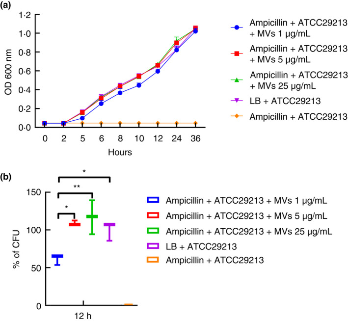
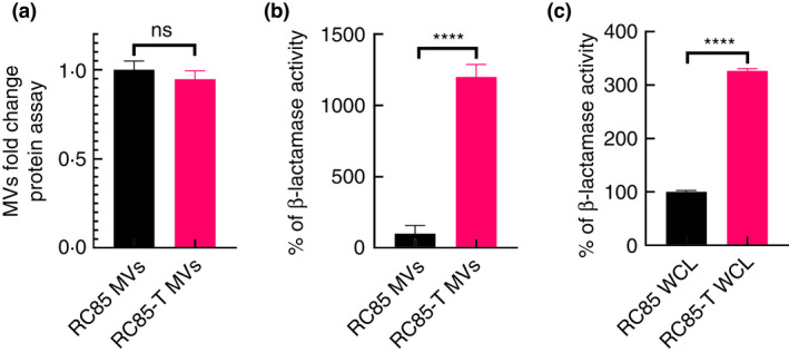
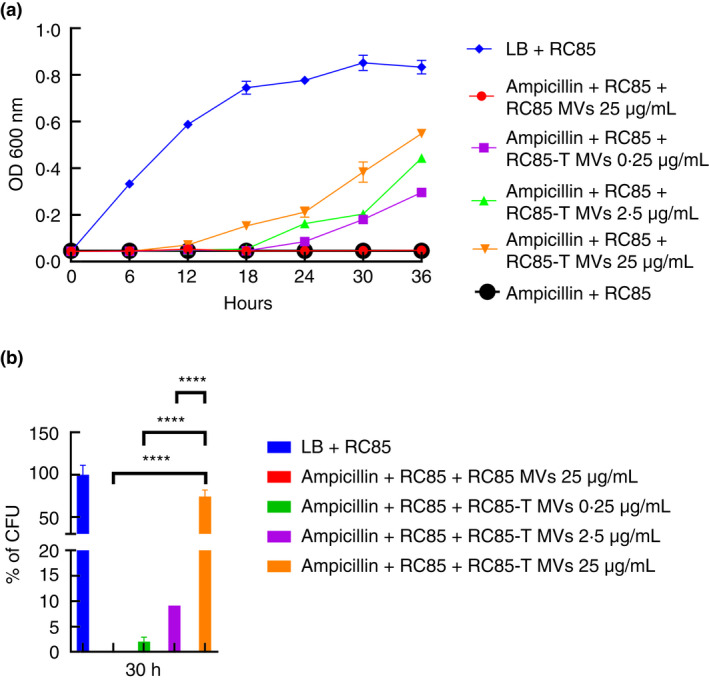
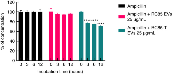
Results: The RC85-T exhibited a higher resistance to β-lactam antibiotics compared to the parent strain. Although the secretion rates of the MVs from RC85-T and the parent strain were nearly equal, the β-lactamase activity of the MVs from RC85-T was 12-times higher than that of MVs from the parent strain, based on equivalent protein concentrations. Moreover, MVs secreted by RC85-T were able to protect β-lactam-susceptible E. coli from β-lactam antibiotic-induced growth inhibition in a dose-dependent manner.

Conclusion: MVs play a role in transferring substances from Gram-positive to Gram-negative bacteria, shown by the release of MVs from RC85-T that were able to protect β-lactam-susceptible bacteria from β-lactam antibiotics.

Significance and impact of study: MVs are involved in the emergence of antibiotic-resistant strains in a mixed bacterial culture, helping us to understand how the spread of multidrug-resistant bacteria could be reduced.

Keywords: antibiotic-resistant bacteria; antibiotic-susceptible bacteria; gram-negative bacteria; gram-positive bacteria; membrane vesicles (MVs); vesicle-mediated transferring of antimicrobial resistance.


Conflict of interest statement

The authors declare no conflict of interest.

Figures

FIGURE 1
FIGURE 1
Physical characterizations of membrane vesicles (MVs) derived from methicillin‐resistant Staphylococcus aureus (MRSA) ST541 cells. Transmission electron microscopy image of MVs derived from ST541 cells (a) (scale bar: 100 nm). The size distribution MVs released from ST541 cells (b), as assessed by dynamic light scattering. Three independent analyses were performed. (c) The zeta potential of MVs from ST541 cells was measured by the zeta‐sizer and each experiment was performed in triplicate
FIGURE 2
FIGURE 2
Membrane vesicles (MVs) from MRSA ST541 can protect β‐lactam‐susceptible Staphylococcus aureus, preventing β‐lactam antibiotic‐induced growth inhibition in the bacteria. (a) Representative growth profiles of β‐lactam‐susceptible S. aureus cells in the presence of growth‐inhibiting concentrations of β‐lactam antibiotics. The growth‐inhibiting concentrations of antibiotics were ampicillin, 60 μg/ml. The data were presented as means and standard error of the mean (SEMs) of at least three independent experiments. (b) The survival percentages of β‐lactam‐susceptible S. aureus cells in the presence of the above‐listed growth‐inhibiting concentrations of antibiotics and MVs were calculated by bacterial counts of cultures at a certain time point (ampicillin, 12 h). CFU of S. aureus cells in medium without any antibiotics were used as a positive control and taken as 100%, to which corresponding CFU of samples were compared. The data were presented as means and SEMs of three independent experiments. *p < 0.05, **p < 0.01
FIGURE 3
FIGURE 3
Percentage of antibiotic‐resistance acquired in RC85 after incubating with membrane vesicles (MVs) treated with and without DNase and representative growth profiles of RC85 and RC85‐T. (a) CFU of cells gaining antibiotic‐resistant in DNase‐treated samples, used as a positive control and taken as 100%, with the corresponding CFU of samples compared to this. The data were presented as means and SEMs of three independent experiments. The abbreviation ‘ns’ means not significant. (b) Growth profiles of respective samples, as obtained by measuring absorbance at 600 nm for up to 10 h every 2 h. The data are presented as means and SEMs of three independent experiments
FIGURE 4
FIGURE 4
Physical characterizations of membrane vesicles (MVs) from RC85‐T and RC85 cells. Transmission electron microscope images of MVs derived from RC85‐T cells (a), and RC85 cells (b) (scale bar: 100 nm); the size distribution of MVs released by RC85‐T cells (c), and RC85 cells (d), as assessed by the zeta‐sizer (three independent analyses were performed;); the zeta potential of MVs released by RC85‐T cells (e), and RC85 cells (f) were measured by the zeta‐sizer and each experiment was performed in triplicate; equal amount of whole cell lysates (WCLs), MVs from RC85‐T and RC85 (g) cells were separated by 10% SDS‐PAGE and then the protein profiles were visualized using Coomassie staining
FIGURE 5
FIGURE 5
Production of membrane vesicles (MVs) isolated from RC85‐T and RC85 cells and differences in β‐lactamase activity between whole cell lysates (WCLs) and MVs. (a) Protein quantification of MVs was conducted using a BCA protein assay. The protein concentration was averaged and normalized to untreated controls to adjust fold change. Data represent means ± SEMs of three independent experiments. The abbreviation ‘ns’ means not significant. (b) β‐lactamase activity of MVs from RC85‐T cells and RC85 cells. (c) β‐lactamase activity of WCLs from RC85‐T and RC85 cells. Respective samples, as obtained by measuring absorbance at 490 nm. The data are presented as means and SEMs of three independent experiments. ****p < 0.0001
FIGURE 6
FIGURE 6
Membrane vesicles (MVs) from RC85‐T cells can protect β‐lactam‐susceptible RC85 from ampicillin‐induced growth inhibition. The growth‐inhibiting concentration of ampicillin was 32 μg/ml. The data represents means and SEMs for at least three independent experiments. (a) the representative growth profiles of β‐lactam‐susceptible RC85 cells in the presence of growth‐inhibiting concentrations of ampicillin and MVs were calculated from bacterial counts of cultures (CFU‐ colony forming units) taken at different time points (ampicillin, 30 h). (b) CFU of RC85 cells in medium without any antibiotics were used as a positive control and taken as 100%, from which the corresponding CFU were determined. The data were presented as means and SEMs of three independent experiments. ****p < 0.0001
FIGURE 7
FIGURE 7
LC‐QQQ‐based assessment of ampicillin concentration following incubation with RC85‐T or RC85 membrane vesicles (MVs) in a cell‐free system. The initial concentration of ampicillin was 20 μg/ml. Twenty‐five micrograms per millilitre of MVs of RC85‐T and RC85 in sterilized 1× PBS were mixed with ampicillin. Filtered 1× PBS containing the ampicillin without MVs were used as a positive control and taken as 100% antibiotic concentration, from which the corresponding concentrations of antibiotic in samples treated with RC85‐T MVs or RC85 MVs were analysed. The concentrations of antibiotics were recorded in triplicate at different time points. Bars indicate standard deviations. **** p < 0.0001

References

    1. Andreoni, F. , Toyofuku, M. , Menzi, C. , Kalawong, R. , Mairpady, S.S. , François, P. et al. (2019) Antibiotics stimulate formation of vesicles in Staphylococcus aureus in both phage‐dependent and‐independent fashions and via different routes. Antimicrobial Agents and Chemotherapy, 63, e01439–18. - PMC - PubMed
    1. Andrews, J.M. (2001) Determination of minimum inhibitory concentrations. The Journal of Antimicrobial Chemotherapy, 48, 5–16. - PubMed
    1. Brown, L. , Wolf, J.M. , Prados‐Rosales, R. & Casadevall, A. (2015) Through the wall: extracellular vesicles in gram‐positive bacteria, mycobacteria and fungi. Nature Reviews. Microbiology, 13, 620–630. - PMC - PubMed
    1. Centers for Disease Control and Prevention . (2013) Antibiotic resistance threats in the United States, 2013. Atlanta, GA: CDC. Available online: https://www.cdc.gov/drugresistance/threat‐report‐2013/pdf/arthreats‐2013.... .
    1. Chopra, I. & Roberts, M. (2001) Tetracycline antibiotics: mode of action, applications, molecular biology, and epidemiology of bacterial resistance. Microbiology and Molecular Biology Reviews, 65, 232–260. - PMC - PubMed

MeSH terms

Substances