. 2020 Dec 21;3(12):8978-8988.
doi: 10.1021/acsabm.0c01229. Epub 2020 Dec 2.

Regulation of Lipoprotein Homeostasis by Self-Assembling Peptides

Affiliations

Regulation of Lipoprotein Homeostasis by Self-Assembling Peptides

Victoria Harbour et al. ACS Appl Bio Mater. .

Abstract

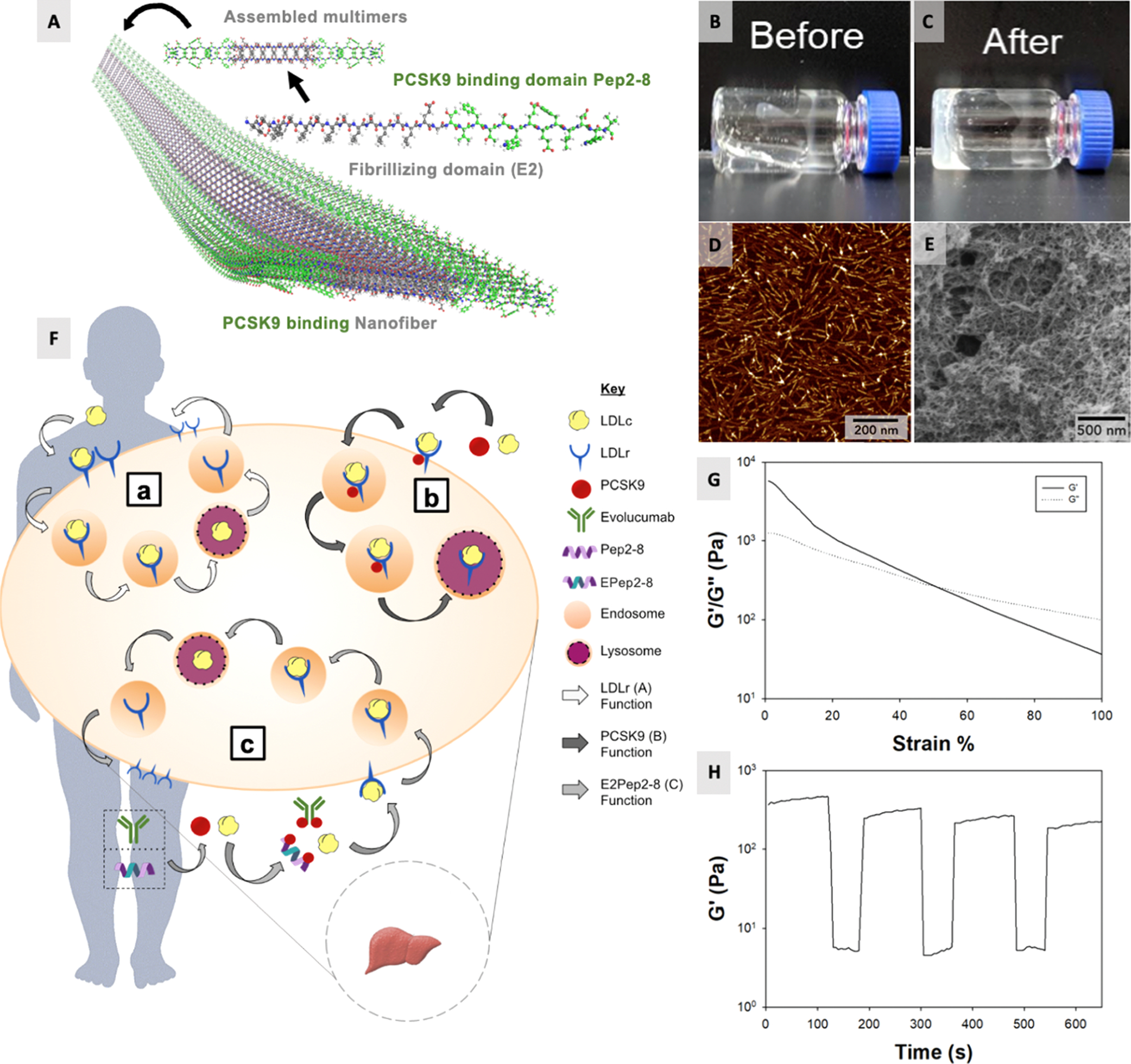
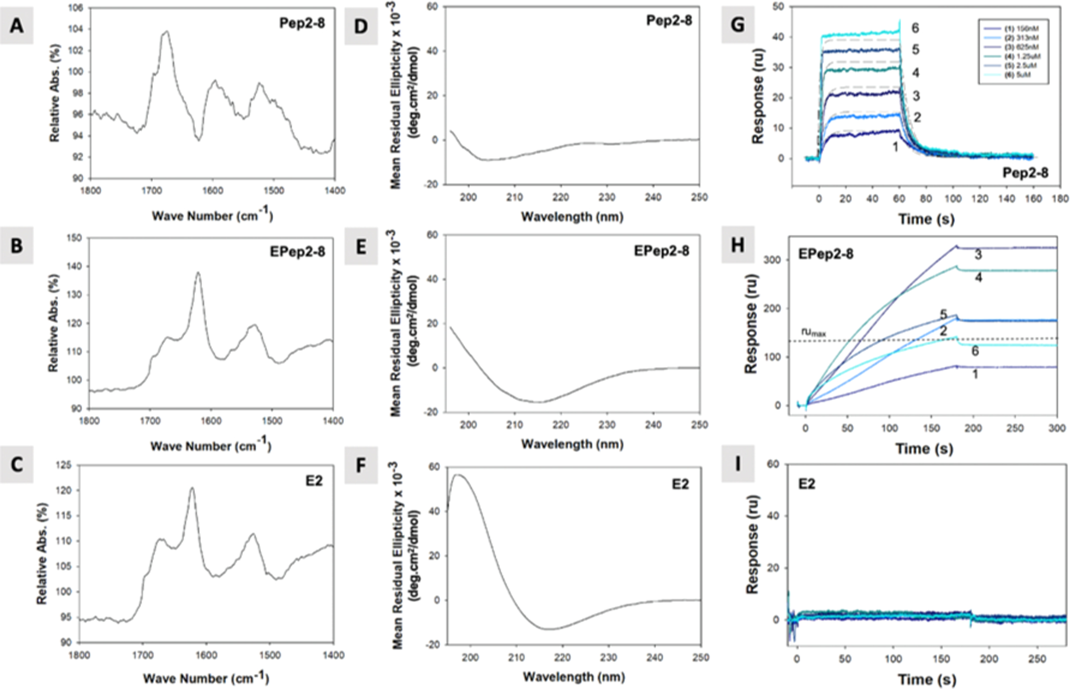
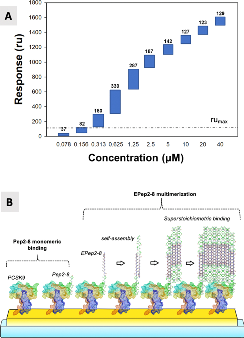
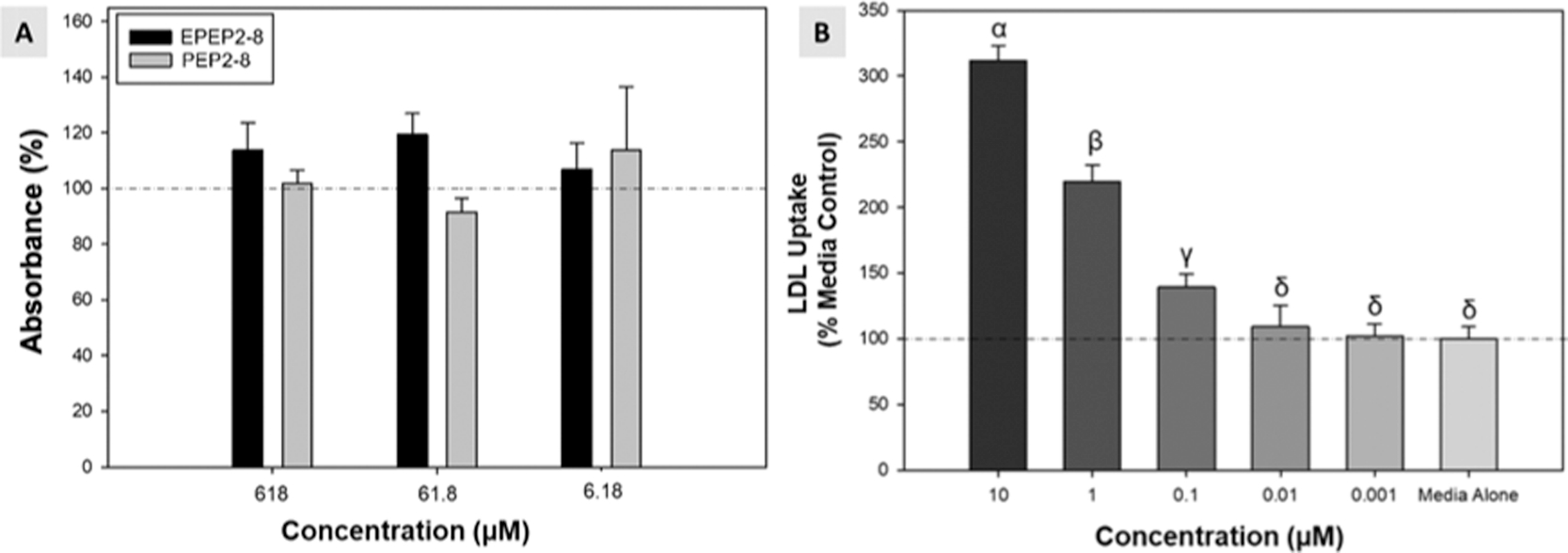
High levels of serum low-density lipoprotein (LDL) cholesterol contribute to atherosclerosis, a key risk factor of cardiovascular diseases. PCSK9 is a circulatory enzyme that downregulates expression of hepatic LDL receptors, concomitantly increasing serum LDL-C. This work investigates a small, self-assembling peptide, EPep2-8, as a peptide inhibitor of PCSK9. EPep2-8 is a multidomain peptide comprising a self-assembling domain, E2, conjugated to a bioactive domain, Pep2-8, previously shown to inhibit PCSK9. The E2 domain facilitates self-assembly of EPep2-8 into long, nanofibrous polymers with an underlying supramolecular β-sheet secondary structure. Intermolecular interactions between nanofibers drive EPep2-8 to form a thixotropic and cytocompatible hydrogel in aqueous and charge-neutral solutions. These properties enable EPep2-8 to be delivered as an in situ depot for regulation of lipoprotein homeostasis. In surface plasmon resonance studies, EPep2-8 bound specifically to PCSK9 with an apparent, noncovalent, and irreversible dissociation, significantly improving the binding affinity of Pep2-8 alone (KD = 667 ± 48 nM). Increased binding affinity of EPep2-8 is primarily due to the superstoichiometric interaction of the peptide with PCSK9. Promisingly, EPep2-8 retains bioactivity in vitro, engendering dose-dependent uptake of LDL-C in hepatocytes. This mechanism of self-assembly on a target site may be a simple method to improve the affinity of peptide inhibitors.

Keywords: LDL; PCSK9; cholesterol metabolism; peptide amphiphiles; self-assembly.


Conflict of interest statement

The authors declare the following competing financial interest(s): The technologies described herein are being investigated for potential commercialization by VK.

Complete contact information is available at: https://pubs.acs.org/10.1021/acsabm.0c01229

Figures

Figure 1.
Figure 1.
SAPs as PCSK9 Inhibitors. (A) EPep2–8 is a SAP containing a fibrillizing domain, E2 (gray carbons), and an appended PCSK9 binding domain, Pep2–8 (green carbons). Multimers assemble into long nanofibers that entangle into hydrogels at concentrations above 0.5 wt %. (B) Peptides form viscous, aqueous solutions and robust, injectable hydrogels upon the addition of equimolar (charge balancing) calcium (C). (D) Tapping mode AFM of EPep2–8 shows a nanofibrillar structure with fiber widths 9.56 nm (±1.21 nm) and heights of 2.205 nm (±0.22 nm). (E) SEM imaging of the critical point-dried EPep2–8 (>1 mg/mL) confirms the nanofibrillar structure of the entangled network of fibers. (F) (a) Hepatocyte LDLR binds LDLc; the complex is endocytosed, and LDLR is recycled to the hepatocyte surface; (b) unless endogenous PCSK9 binds LDLR and induces degradation of the entire complex, diminishing surface LDLR and cholesterol metabolism; (c) subcutaneously or IV-administered PCSK9 inhibitors, such as evolocumab, Pep2–8, and our EPep2–8, can effectively bind and inhibit the PCSK9 function. (G) Viscoelastic hydrogels >1 wt % exhibit a thixotropic behavior; at low shear strain (1%), hydrogels exhibit high storage moduli (>100 Pa) and at high shear strain (100%), hydrogels exhibit low shear storage moduli (~10 Pa); an inverse trend was seen with G″. (H) Little to no hysteresis was observed after repeated low and high strain loading, demonstrating facile injectability as in situ depots.
Figure 2.
Figure 2.
Chemical characterization and binding of peptides. (A–C) ATR–FTIR of peptides shows a disordered secondary structure for Pep2–8 (A) and characteristic peaks at (~1622 cm−1) for self-assembling constructs, indicating the β-sheet-specific amide-1 secondary structure between EPep2–8 (B) and E2 (C) nanofibers. (D–F) CD spectra of peptide structures indicate a secondary structure. (D) MRE values at ~215 nm are consistent with the disorganized secondary structure for the non-SAP, Pep2–8. (E,F) SAPs (EPep2–8, E2) contain characteristic MRE minima at ~215 nm and increase at ~195 nm, consistent with the β-sheet secondary structure. (G–I) Sensogram of binding to an immobilized PCSK9 surface. Binding kinetics were assessed by SPR at a selected range of analyte concentrations (curve 1: 156 nM to curve 6: 5 μM, twofold dilutions). The decrease in ru for concentration 4, 5, and 6 is due to saturation of the biotin-PCSK9 binding site. (G) Pep2–8 binding kinetics to immobilized PCSK9 (ka = 2.09 × 10⁵ M−1 s−1, kd = 0.151 s−1, and KD = 721 ± 0.9 nM). (H) EPep2–8 shows apparent irreversible binding at all concentrations. (I) Control peptide E2 shows no binding to the PCSK9 surface.
Figure 3.
Figure 3.
Superstoichiometric binding. (A) EPep2–8 accumulation on a PCSK9 bound surface. Values above each bar represents the maximum ru binding achieved during the injection. The dotted line, rumax, indicates the theoretical maximum binding for a 1:1, PCSK9/EPep2–8 interaction (135 ru). (B) EPep2–8 may self-assemble on immobilized PCSK9, giving rise to superstoichiometric binding (>135 ru) and greater binding affinity between EPep2–8 and PCSK9.
Figure 4.
Figure 4.
Cytocompatibility and efficacy of novel peptide conjugates. (A) CCK-8 (Dojindo) metabolic activity in fibroblast cells was measured by fluorescence to assess cytocompatibility of Pep2–8 and EPep2–8. Normalized to a medium-only control, no conditions exhibited cytotoxicity (p > 0.05). (B) Hepatocytes incubated with Bodipy fluorescent LDL show dose-dependent uptake of LDL in response to increasing levels of EPep2–8. Greek letters denote groups with statistically significant difference (p < 0.05).

References

    1. Khan T Cardiovascular Diseases. https://www.who.int/health-topics/cardiovascular-diseases/#tab=tab_1 (accessed June 22, 2020).
    1. Cardiovascular Disease: Data and statistics. https://www.euro.who.int/en/health-topics/noncommunicable-diseases/cardi... (accessed June 17, 2020).
    1. Hansson GK Inflammation, Atherosclerosis, and Coronary Artery Disease. N. Engl. J. Med. 2005, 352, 1685–1695. - PubMed
    1. Libby P; Theroux P Pathophysiology of Coronary Artery Disease. Circulation 2005, 111, 3481–3488. - PubMed
    1. Cannon B Cardiovascular disease: Biochemistry to behaviour. Nature 2013, 493, S2–S3. - PubMed

LinkOut - more resources