. 2022 Feb 23;10(1):e0099121.
doi: 10.1128/spectrum.00991-21. Epub 2022 Jan 12.

The Mechanism of Action of Ginkgolic Acid (15:1) against Gram-Positive Bacteria Involves Cross Talk with Iron Homeostasis

Affiliations

The Mechanism of Action of Ginkgolic Acid (15:1) against Gram-Positive Bacteria Involves Cross Talk with Iron Homeostasis

Zewen Wen et al. Microbiol Spectr. .

Abstract

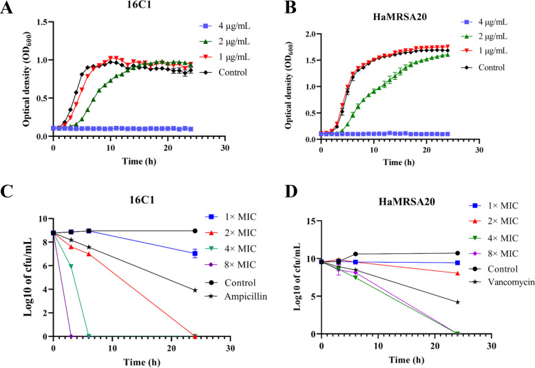
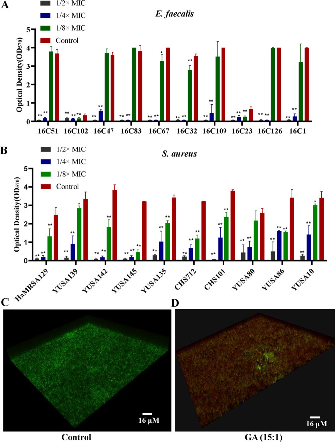
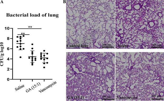
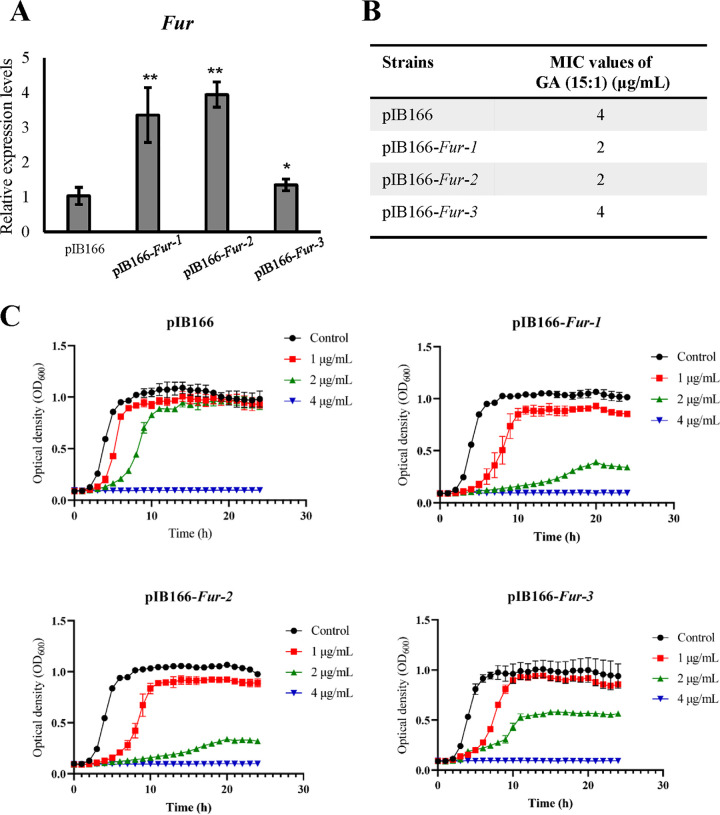
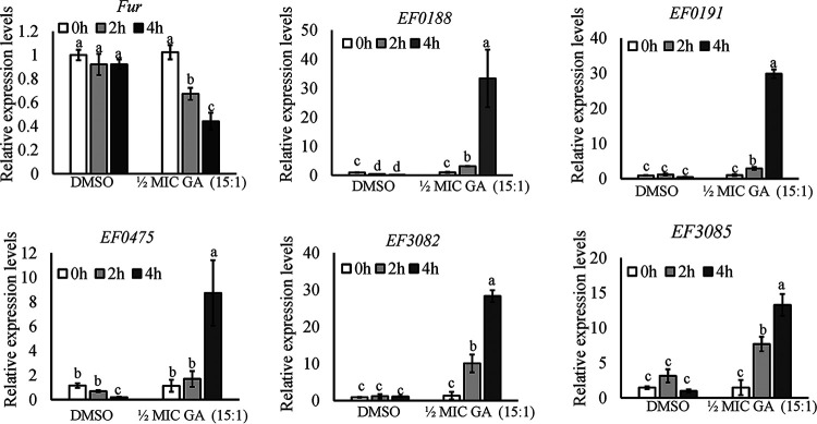
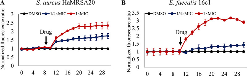
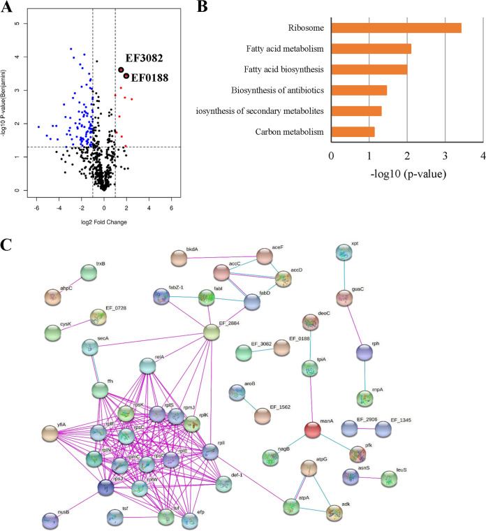
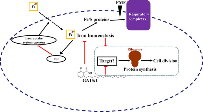
With the increasing reports of community-acquired and nosocomial infection caused by multidrug-resistant Gram-positive pathogens, there is an urgent need to develop new antimicrobial agents with novel antibacterial mechanisms. Here, we investigated the antibacterial activity of the natural product ginkgolic acid (GA) (15:1), derived from Ginkgo biloba, and its potential mode of action against the Gram-positive bacteria Enterococcus faecalis and Staphylococcus aureus. The MIC values of GA (15:1) against clinical E. faecalis and S. aureus isolates from China were ≤4 and ≤8 μg/mL, respectively, from our test results. Moreover, GA (15:1) displayed high efficiency in biofilm formation inhibition and bactericidal activity against E. faecalis and S. aureus. During its inhibition of the planktonic bacteria, the antibacterial activity of GA (15:1) was significantly improved under the condition of abolishing iron homeostasis. When iron homeostasis was abolished, inhibition of planktonic bacteria by GA (15:1) was significantly improved. This phenomenon can be interpreted as showing that iron homeostasis disruption facilitated the disruption of the functions of ribosome and protein synthesis by GA (15:1), resulting in inhibition of bacterial growth and cell death. Genetic mutation of ferric uptake regulator (Fur) led to GA (15:1) tolerance in in vitro-induced resistant derivatives, while overexpression of Fur led to increased GA (15:1) susceptibility. Additionally, GA (15:1) significantly decreased the bacterial loads of S. aureus strain USA300 in the lung tissues of mice in a pneumonic murine model. Conclusively, this study revealed an antimicrobial mechanism of GA (15:1) involving cross talk with iron homeostasis against Gram-positive pathogens. In the future, the natural product GA (15:1) might be applied to combat infections caused by Gram-positive pathogens. IMPORTANCE The increasing emergence of infectious diseases associated with multidrug-resistant Gram-positive pathogens has raised the urgent need to develop novel antibiotics. GA (15:1) is a natural product derived from Ginkgo biloba and possesses a wide range of bioactivities, including antimicrobial activity. However, its antibacterial mechanisms remain unclear. Our current study found that the function of ferric uptake regulator (Fur) was highly correlated with the antimicrobial activity of GA (15:1) against E. faecalis and that the antibacterial activity of GA (15:1) could be strengthened by the disruption of iron homeostasis. This study provided important insight into the mode of action of GA (15:1) against Gram-positive bacteria and suggested that GA (15:1) holds the potential to be an antimicrobial treatment option for infection caused by multidrug-resistant Gram-positive pathogens.

Keywords: E. faecalis; Fur; antibacterial mechanism; ferric uptake regulator; ginkgolic acid (15:1); iron homeostasis.


Conflict of interest statement

The authors declare no conflict of interest.

Figures

FIG 1
FIG 1
Bacterial growth curve and bactericidal effect analysis of GA (15:1) against E. faecalis (16C1) and MRSA HaMRSA20. (A, B) Impact of GA (15:1) at different concentrations (1/4×, 1/2×, and 1× MIC) on the bacterial growth of vancomycin-intermediate E. faecalis 16C1 and MRSA HaMRSA20 planktonic cells. (C, D) Time-kill assay of GA (15:1) with 1×, 2×, 4×, and 8× MIC against vancomycin-intermediate E. faecalis isolate 16C1 and MRSA HaMRSA20 cells at exponential phase. Data are presented as mean values ± standard deviations (SD). The control concentration (for both ampicillin and vancomycin) was 4× MIC.
FIG 2
FIG 2
Antibiofilm activity of GA (15:1) against S. aureus and E. faecalis. (A, B) Significant inhibition of the biofilm formation of E. faecalis (A) and S. aureus (B) by GA (15:1) at different subinhibitory concentrations. The 10 E. faecalis strains, 5 MSSA strains, and 5 MRSA strains were treated with GA (15:1) at 1/2×, 1/4×, and 1/8× MIC for 24 h, and the biofilm formation was determined by crystal violet staining. The data presented are the average values from three independent experiments (means ± SD). P values are for comparison with the control: *, P < 0.05; **, P < 0.01 (Student’s t test). (C, D) Effect of GA (15:1) at 8× MIC against the viable cells embedded in mature biofilm of E. faecalis isolate 16C102. Bacterial cells were inoculated onto 96-well polystyrene microtiter plates for 24 h at 37°C until mature biofilms were formed. After being treated with GA (15:1) at 8× MIC or solvent control for another 24 h, the viability of the cells embedded in the mature biofilm was observed by confocal microscopy using LIVE/DEAD staining.
FIG 3
FIG 3
Protective effect of GA (15:1) against S. aureus USA300 pneumonia. Female BALB/c mice (n = 12/group) were challenged with S. aureus USA300 by nasal drip; GA (15:1) (25 mg/kg) or vancomycin (25 mg/kg) was administered to the mice by intraperitoneal injection 2 h before the bacterial challenge. (A) Bacterial burdens in the lungs were determined at 24 h postinfection. Data are presented as the mean values ± SD. **, P < 0.01 (Student’s t test). (B) Hematoxylin and eosin (H&E) staining (10×) of lung tissue showed that histopathological change (inflammatory cells) was significantly reduced after GA (15:1) treatment. Scale bars, 300 μm.
FIG 4
FIG 4
fur transcript levels and susceptibility analysis of GA (15:1) in three independent Fur overexpression transgenic isolates and pIB166 empty-vector control in E. faecalis strain OG1RF. (A) Three independent transgenic isolates of Fur overexpression strains (pIB166-fur-1, pIB166-fur-2, and pIB166-fur-1) and their empty-vector control in the exponential phase were used for total RNA extraction, and qRT-PCR was performed to determine the fur transcript levels. gdh was used as an internal control. Data are presented as the mean values ± SD. *, P < 0.05; **, P < 0.01 (Student’s t test). (B) MICs of GA (15:1) against Fur overexpression transgenic isolates and pIB166 empty-vector control in E. faecalis. (C) Bacterial growth curves of Fur overexpression E. faecalis strains after exposure to GA (15:1). Data are presented as mean values ± SD.
FIG 5
FIG 5
GA (15:1) alters the transcript levels of fur and Fur-regulated operons in E. faecalis. qRT-PCR was used to determine the expression levels of fur, EF0188, EF0191, EF0475, EF3082, and EF3085 in E. faecalis strain OG1RF at different time points after treatment with 1/2× MIC of GA (15:1). gdh was used as an internal control. Data are mean values ± SD from three technical replicates. Different letters indicate statistically significant differences based on ANOVA (P < 0.05).
FIG 6
FIG 6
GA (15:1) rapidly dissipated the proton motive force of S. aureus and E. faecalis cells. S. aureus HaMRSA20 (A) and E. faecalis 16C1 (B) cells were treated with DiBAC4(3) for 10 min and then treated with 1/4× or 1× MIC GA (15:1) for 20 min. The fluorescence of DiBAC4(3) was excited at 492 nm with an emission at 518 nm. Data were normalized to the values of the DMSO-treated control cells.
FIG 7
FIG 7
Differential expression of proteins between the control groups and GA (15:1)-treated groups. (A) Volcano plots show log2 fold changes of protein levels after treatment of E. faecalis OG1RF cells with GA (15:1) (2 μg/mL, 1/2× MIC) compared to DMSO treatment. Blue dots represent proteins whose expression has been found to be inhibited by GA (15:1). Red dots represent proteins that are upregulated by GA (15:1). Black circles represent ABC transporters of iron. Data represent average values, and P values were calculated using a 2-sided 2-sample t test; n = 3 independent experiments per group. (B) KEGG Pathway terms of the differentially expressed proteins between the two groups. (C) Protein-protein interaction network analysis for proteins differentially expressed between the control groups and GA (15:1)-treated groups. Each node represents a protein, and each edge represents an interaction between proteins. Only known interactions were included. Disconnected nodes are hidden.
FIG 8
FIG 8
Hypothetical model for the antibacterial mode of action of GA (15:1) against Gram-positive pathogens by cross talk with iron homeostasis. When GA (15:1) is present, it destabilizes the iron homeostasis and creates a state of iron starvation, which triggers the iron starvation response: Fur-mediated repression of iron uptake transporters is relieved, and iron transporters are upregulated, allowing Fe3+ to enter the bacterial cells. In addition, GA (15:1) blocks protein biosynthesis and inhibits the growth and cell division of the bacterium through unidentified targets. Meanwhile, the iron starvation condition can strengthen this process.

References

    1. Leclercq R. 2009. Epidemiological and resistance issues in multidrug-resistant staphylococci and enterococci. Clin Microbiol Infect 15:224–231. doi:10.1111/j.1469-0691.2009.02739.x. - DOI - PubMed
    1. Woodford N, Livermore DM. 2009. Infections caused by Gram-positive bacteria: a review of the global challenge. J Infect 59(Suppl 1):S4–S16. doi:10.1016/S0163-4453(09)60003-7. - DOI - PubMed
    1. D’Costa VM, King CE, Kalan L, Morar M, Sung WW, Schwarz C, Froese D, Zazula G, Calmels F, Debruyne R, Golding GB, Poinar HN, Wright GD. 2011. Antibiotic resistance is ancient. Nature 477:457–461. doi:10.1038/nature10388. - DOI - PubMed
    1. Inagaki K, Lucar J, Blackshear C, Hobbs CV. 2019. Methicillin-susceptible and methicillin-resistant Staphylococcus aureus bacteremia: nationwide estimates of 30-day readmission, in-hospital mortality, length of stay, and cost in the United States. Clin Infect Dis 69:2112–2118. doi:10.1093/cid/ciz123. - DOI - PubMed
    1. Beganovic M, Luther MK, Rice LB, Arias CA, Rybak MJ, LaPlante KL. 2018. A review of combination antimicrobial therapy for enterococcus faecalis bloodstream infections and infective endocarditis. Clin Infect Dis 67:303–309. doi:10.1093/cid/ciy064. - DOI - PMC - PubMed

Publication types

MeSH terms